- Research
- Open access
- Published:
Value of a combined magnetic resonance-enhanced and diffusion-weighted imaging dual-sequence radiomics model in predicting the efficacy of high-intensity focused ultrasound ablation for uterine fibroids
BMC Medical Imaging volume 25, Article number: 53 (2025)
Abstract
Objective
To establish a joint radiomics model based on T1 contrast-enhanced (T1C) imaging and diffusion-weighted imaging (DWI), and investigate its value in predicting the efficacy of high-intensity focused ultrasound (HIFU) in ablating uterine fibroids.
Methods
This multicenter retrospective study included 195 patients with uterine fibroids. Their data were divided into training (n = 120), internal test (n = 30), and external test (n = 45) sets. The radiomic features were extracted from T1C and DWI sequences. Logistic regression was used to develop the T1C, DWI, integration, and joint models, and receiver operating characteristic curves were used to assess model performance. The Delong test was used to compare the predictive efficacies of different models, and the best model was used for external validation and development of the nomogram.
Results
Eight T1C features, six DWI features, and three imaging features were retained for the modeling. The areas under the curve were 0.852 and 0.769 for the integrated model on the training and internal test sets, respectively; 0.857 and 0.824 for the joint model on the training and internal test sets, respectively, which were higher than those of the single-sequence model; and 0.857 for the joint model on the external test set.
Conclusions
A joint radiomics model based on T1C and DWI data can effectively predict the efficacy of HIFU for ablating uterine fibroids and guide the development of individualized clinical treatment plans.
Introduction
Uterine fibroids are benign tumors of the smooth muscle of the uterus, with a prevalence of approximately 20–40% in women of childbearing age [1]. They may be asymptomatic, but 20–50% of patients experience abnormal uterine bleeding, menstrual disorders, pain, and reduced fertility, which markedly affects the quality of life [1,2,3,4,5].
Treatment options for uterine fibroids include surgery, medication, and other minimally or non-invasive treatments. The radical surgery for uterine fibroids is total hysterectomy, but this is not suitable for patients with reproductive needs. Minimally invasive surgeries are required for patients with reproductive needs, but such surgeries are more demanding on the operator and are associated with longer recovery durations [6]. High-intensity focused ultrasound (HIFU) relies on the thermal and mechanical energy of ultrasonic waves to cause coagulative necrosis of tissues under the guidance of ultrasound (USgHIFU) or MRI (MR-HIFU). This method is safe and effective and can significantly improve the clinical symptoms of patients. It may be an alternative to surgical treatment [7], complementing existing treatment options. Non-perfused volume ratio (NPVR) after HIFU for uterine fibroids is an important index for evaluating the ablation effect [8]. Patients with higher NPVR have better clinical efficacy and lower probability of recurrence [9, 10]. However, not all patients with fibroids are suitable for HIFU treatment [11], so the preoperative prediction of NPVR will be helpful in patient selection. Radiomics can convert medical images into high-throughput data, which can be useful for discovering subtle differences in the images that cannot be recognized by the human eye. This information can be used to achieve assisted diagnosis or prediction [12–13].
Previous studies have shown that the degree of T1 contrast-enhanced imaging (T1C) enhancement is a factor closely related to the efficacy of ablation, and higher degrees of enhancement are associated with lower NPVR [14]; diffusion-weighted imaging (DWI) reflects the perfusion, and the NPVR is correlated with the increase in DWI signal intensity [15]. Models developed in previous studies have been predominantly based on single sequences or T2W combined with T1C sequences, and DWI sequences have predominantly yielded features from apparent diffusion coefficient (ADC) images [16–17]. Therefore, DWI sequences have some research value. However, few studies have combined T1C and DWI sequences.
The aim of this study was to investigate the value of a joint radiomics model based on T1C and DWI data in predicting the efficacy of HIFU ablation of uterine fibroids to guide the clinical selection of patients. We present this paper based on a list of CLEAR and METRICS reports (Supplementary file 1).
Materials and methods
Patients
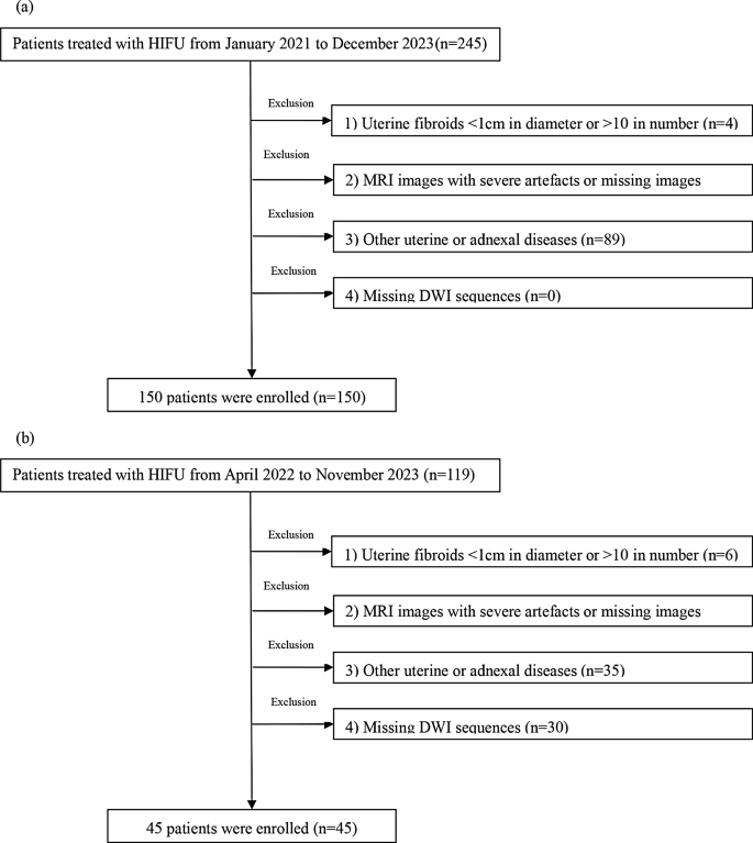
This study involved patients who received USgHIFU treatment from January 2021 to December 2023 at Yongchuan Hospital of Chongqing Medical University (our hospital) and the Second Affiliated Hospital of Chongqing Medical University (the other hospital).The inclusion criteria were as follows: (a) age of > 18 years and non-menopausal; (b) the pelvic MRI was conducted within one week before and one week after the HIFU treatment,, with scanning sequences including T1C and DWI sequences; (c) uterine fibroids diagnosed based on clinical symptoms (such as abnormal uterine bleeding, change in menstruation, pain, or infertility) without any prior treatment; and (d) clear MRI images. The exclusion criteria were as follows: (a) uterine fibroid diameter of < 1 cm or number of > 10, (b) severe artifacts or missing MRI images, (c) coexistence with other uterine or adnexal diseases, and (d) lack of DWI sequences (b-value of 50 s/mm2). The patient selection process is shown in Fig. 1a, b.
Patient grouping
Previous studies have shown that patients with NPVR < 70% have an increased risk of clinical reintervention [18]. Meanwhile, the volume of residual tumor tissue in leiomyomas with NPVR > 70% shows a significant trend of reduction within 12 months after surgery [18]. The participants in this study were divided into the sufficient (NPVR > 70%) and non-sufficient (NPVR ≤ 70%) ablation groups with NPVR of 70% as the threshold. Their data were randomly divided into the training (n = 120) and internal test (n = 30) sets at a ratio of 8:2, while the data of the patients in the Second Affiliated Hospital of Chongqing Medical University were allocated to the external test set (n = 45) (See Supplementary file 3 for sample size calculations). The ellipsoidal volume was calculated using the following formula [19]:
where Da is anterior-posterior diameter, Dl is left-right diameter, and Du is up-and-down diameter.
The diameters were measured from the preoperative T2WI and postoperative T1C images to calculate the uterine fibroid volume V1 and non-perfused area volume NPV. The NPVR was calculated as follows:
The member of our team who is responsible for data collection is in charge of calculating NPVR.
Magnetic resonance examination methods
All patients underwent T1C and DWI before treatment. The contrast agent was injected intravenously at a dose of 0.2 ml/kg and a flow rate of 2.0–2.5 ml/s. The delayed-phase images were acquired 2 min after injection of the contrast agent. Siemens Verio dot 3.0T MR was used in our hospital. Siemens Prisma 3.0T and Siemens Avanto 1.5T MR were used in other hospitals. The scanning parameters are shown in supplementary table S1.
Imaging features and clinical features
The patient data collected included age, fibroid volume, subcutaneous fat thickness, shortest distance from the center of the target fibroid to the skin at the ventral level, location of fibroid, degree of enhancement on T1WI [3,4,5] (below the myometrium = mild, similar to the myometrium = moderate, above the myometrium = significant) (Supplementary figure S1), and intensity of the signal on DWI (with reference to the myometrium, below the myometrium = low signal, similar to the myometrium = medium signal, above the myometrium = high signal) (Supplementary figure S2).
Image segmentation
Two radiology abdominopelvic team physicians with five or more years of experience manually outlined the entire uterine fibroids to create a region of interest (ROI), no other individuals participated in this task. They selected the preoperative T1C (delayed-phase) and DWI (b-value of 50s/mm2) sequences of the uterine fibroids and discarded the images with poor edge contours to prevent the influence of the volumetric effect (they were blinded to the postoperative scan results throughout the process). To assess the reproducibility of the radiomics features, the images of 50 patients were randomly selected from the T1C and DWI sequences of all the uterine fibroids after one month. The ROIs were outlined again by the two radiologists mentioned above. Interclass correlation coefficients (ICC) were used to evaluate the consistency of the indicators; an ICC of > 0.75 was considered good.
Screening of radiomics features
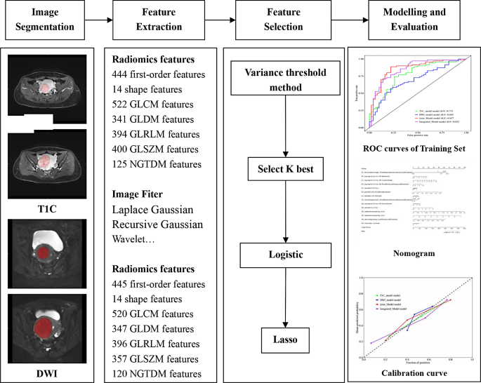
In the image preprocessing stage, we set the binWidth to 25 and resampled the image using BSpline interpolation. The image was resampled to a voxel size of 1 × 1 × 1 mm. The original image was transformed using various filters, such as Laplace Gaussian, Recursive Gaussian, and wavelet transform filtering. We were using Pyradiomics extracted radiomics features and we followed international standards throughout the process. The radiomic features were extracted from the outlined ROIs, and those with ICCs of > 0.75 were selected. Z-score normalization was first used to eliminate the quantitative differences between the radiomics features, followed by the variance thresholding method (the threshold was set to 0.8 to remove features with variance less than 0.8), the best K selection (to remove features that are not significantly different between the two groups), logistic regression, and the least absolute shrinkage and selection operator (LASSO) were used for feature screening. The Radiomics feature parameter has been set to its default value.
Model building
The radiomic features retained after dimensionality reduction and those with statistically significant differences between the two groups were used to establish the T1C model, DWI model, integrated model (joint model and imaging features), and joint model (T1C and DWI) using logistic regression. The predictive values of the models were evaluated using the area under the curve (AUC), sensitivity, specificity, accuracy, and precision. The differences between the AUCs of the models were determined using the Delong’s test. The model with the best predictive performance was used for external validation and development of the nomogram. The above processes were carried out using the uAI Research Portal (V730) software. A flowchart of the process for the development of the radiomics model is shown in Fig. 2.
Statistical analysis
SPSS 26.0 statistical analysis software was used for the statistical analysis. Measurement data conforming to the normal distribution are expressed as mean ± standard deviation, and the t-test was used to compare the groups. The measurement data not conforming to the normal distribution are expressed as median (interquartile range), and Wilcoxon’s rank sum test was used to compare them. The chi-squared or Fisher’s exact test was used to compare the counting data. P < 0.05 was considered statistically significant.
Results
General information
Two hundred and forty-five patients received HIFU treatment between January 2021 and December 2023 at Yongchuan Hospital of Chongqing Medical University (our hospital). Based on the inclusion and exclusion criteria, 150 patients were included from our hospital. Their mean age was 45.1 ± 6.4 (23–59) years. Forty-five patients were included from the Second Affiliated Hospital of Chongqing Medical University (the other hospital). Their mean age was 38.5 ± 7.5 (22–53) years. The general patient information is shown in Table 1. The sufficient and non-sufficient ablation groups had statistically significant differences related to age, shortest distance from the center of the target fibroid to the skin at the ventral level, degree of T1WI enhancement, and DWI signal intensity (P < 0.05). The differences in the degree of T1WI enhancement between the sufficient and non-sufficient ablation groups in the external test set were statistically significant (P < 0.05).
Radiomics characterization
From the T1C and DWI sequences, 2228 and 2199 radiomic features were extracted, respectively, and classified into first-order, shape, and texture features. Eight and six features were retained in the T1C and DWI sequences after screening using the variance threshold, select K best, logistic regression, and LASSO (supplementary table S2).
Modeling and evaluation
The prediction efficacy of each model is shown in Table 2. The AUC of the joint model was 0.857, and those of the joint models for the internal and external test sets were 0.824 and 0.857, respectively (Fig. 3). After Delong’s test, the AUCs of the joint and integrated models were higher than those of the T1C and DWI models, and the differences between the AUCs of the joint and integrated models and those of the T1C and DWI models for the training set were statistically significant, respectively (P < 0.05). However, the differences in the AUCs for the test set were not statistically significant (P > 0.05), which may be attributed to the small sample size. The difference between the AUC values of the joint and integrated models was not statistically significant (P > 0.05). The results indicated that the joint model had a better predictive value than the individual T1C and DWI models. It also had better predictive ability on the external test set (AUC = 0.857).
A nomogram was developed using the radiomics features of the joint model (Fig. 4a), and the calibration curves (Fig. 4b, c) showed that the predicted results of the combined model were in better agreement with the actual results. The detrended correspondence analysis plot showed that the joint model had the highest net benefit and a wider threshold probability than the other models (Fig. 4d).
Nomograms constructed based on the joint model (a). Calibration curves of the nomogram in the training and validation cohorts (b, c). The black dashed line represents the ideal prediction, and the green, blue, red, and purple lines represent the predictive ability of the T1C model, the DWI model, the joint model, and the integrated model. The closer they are to the dotted line, the higher the prediction accuracy of the model. Decision curve analysis for the nomogram (d). The green, blue, red, and purple lines represent the expected net benefits of the clinical T1C model, the DWI model, the joint model, and the combined model
Discussion
In this study, we developed T1C, DWI, joint, and integrated models using logistic regression. Among them, the joint model (AUC = 0.857) based on eight radiomics features of T1C sequences and six radiomics features of DWI sequences had the best predictive performance. This model had an AUC of 0.857 on the external test, which was superior to that reported by previous studies[20], indicating that the model had a better potential for predicting the efficacy of HIFU during the preoperative period. There were also some differences in the instruments used for the MR examination of our patients and those from other hospitals. However, the external validation results were still better, which further validated the performance of the model. We also used tools such as nomograms and decision curves to evaluate the individual models and showed that the joint model had the highest overall net benefit in predicting ablation rates and had the best clinical utility. This model is expected to guide clinical application, and patients with uterine fibroids can benefit from its use. The flowchart used by clinicians for this model is shown in supplementary figure S3.
Some investigators have used radiomics to predict the outcomes after HIFU for uterine fibroids. Li et al. [14] developed prediction models based on T2WI and T1C sequences using LightGBM and XGBoost, respectively, and the results have shown that the performance of the model based on LightGBM was better (AUC = 0.848), however, they are all single-sequence models. Zheng et al. [17] developed prediction models based on T2WI and DWI sequences using various machine learning classifiers, and the model based on support vector machine performed better (AUC = 0.822), however, this model did not select the commonly used T1C sequence. Jiang et al. [4] developed a prediction model based on the T1C sequence using a support vector machine and obtained satisfactory results (AUC = 0.83), but tools such as nomograms and decision curves were not utilized. The aforementioned studies are all single-center studies and lack external validation. Jacobs et al. [21] showed that T1C sequences determined the activity of the fibroid tissue by the enhancement of the fibroid before ablation. HIFU treatment was feasible if the fibroid was “alive.” DWI was also used to differentiate the viable from non-viable fibroid tissues; after ablation, the enhancement of T1C was markedly reduced, and the signal intensity on DWI increased. There were good agreement and correlation between the two in localizing the area of uterine fibroid treatment; DWI has been used as an adjunct to T1C. Therefore, we analyzed the T1C and DWI sequences and established a dual-sequence combined model. The combination of sequences can reveal more information. Meanwhile, we also evaluate the model’s performance through external validation. Advanced tools such as nomograms and decision curves are employed to assess the clinical practicality of the model. These are the strengths of our model.
The differences in the shortest distance from the center of the target fibroid to the skin at the ventral level, degree of enhancement of the fibroids on T1WI, and signal intensity of DWI between the sufficient and non-sufficient ablation groups of our hospital in this study were statistically significant. HIFU relies on the mechanical and thermal effects of ultrasound to gather energy at the target tissue under the guidance of imaging equipment to cause coagulative necrosis of the target tissue [22]. When the target tissue is farther away, more energy is required because the interfaces in the acoustic pathway cause absorption, reflection, and scattering of ultrasound waves. This leads to increased attenuation of ultrasound energy and a decrease in the NPVR [13, 23]. The signal intensity of T1C reflects the blood supply of the fibroid, and a higher degree of fibroid enhancement is associated with richer blood supply, worse energy aggregation, and greater difficulty of ablation [24]. Previous studies have shown that the DWI of fibroids can reveal perfusion, cellular characteristics, and microcirculatory status [15, 25], and NPVR correlates with an increase in the signal intensity of DWI [15]. However, imaging characteristics are measured artificially, can be influenced by various factors, and are subjective. These may explain the statistical results of the imaging characteristics of the patients in the external test.
The limitations of this study are as follows. First, the sample size is small and limited external validation. Second, this study only extracted the features of the DWI images with low b-values. Third, our model only united two sequences, T1C and DWI, and did not unite sequences such as T2WI. The fusion of T2 sequences can provide some value because more and more comprehensive features are included in the model. However, whether the model will be better or not needs to be verified experimentally. We are prepared to unite T2WI, T1C, DWI, and ADC sequences in the future, and at the same time, expand the sample size as much as possible, with the hope of developing a better performing and more stable model. Fourth, early T1C images were not analyzed for the following reasons. (a) Previous researchers have already studied early T1C images with good results (AUC = 0.83), and we are ready to explore the characteristics of other images. (b) This was a retrospective study, and we found that most of the of the T1C images of the patients were only clearer for the delayed phase. Therefore, we had to select the delayed-phase images, resulting in the inability to perform comparisons of the various phases of T1C. Regarding this shortcoming, we are going to communicate with the technicians to collect images for each period when the subsequent patients are examined.
In conclusion, the joint radiomics model based on the T1C and DWI sequences can effectively predict the efficacy of HIFU ablation of uterine fibroids preoperatively and provide an imaging basis for the selection of treatment plans.
Data availability
Data used to support the findings of this study are available from the corresponding author.
References
Ahmad A, Kumar M, Bhoi NR, et al. Diagnosis and management of uterine fibroids: current trends and future strategies. J Basic Clin Physiol Pharmacol. 2023;34(3): 291–310, https://doiorg.publicaciones.saludcastillayleon.es/10.1515/jbcpp-2022-0219.
Giuliani E, As-Sanie S, Marsh EE. Epidemiology and management of uterine fibroids. Int J Gynaecol Obstet. 2020;149(1): 3–9, https://doiorg.publicaciones.saludcastillayleon.es/10.1002/ijgo.13102.
Li C, Xiao Z, He Z, et al. Value of integrated T2WI and CE-T1WI imaging-based histological models in predicting the efficacy of HIFU ablation of uterine fibroids. Chin J Magn Reson Imaging. 2023;14(6):45–51. https://doiorg.publicaciones.saludcastillayleon.es/10.12015/j.issn.1674-8034.2023.06.007.
Jiang Y, Huang X, Qin S, et al. The value of enhancement-based MRI imaging histology model in predicting the efficacy of HIFU ablation of uterine fibroids. J Clin Rad. 2022;41(11):2095–2100. https://doiorg.publicaciones.saludcastillayleon.es/10.13437/j.cnki.jcr.2022.11.008.
Qin S, Jiang Y, Wang F, et al. Development and validation of a combined model based on dual-sequence MRI radiomics for predicting the efficacy of high-intensity focused ultrasound ablation for hysteromyoma. Int J Hyperthermia. 2023;40(1): 2149862, https://doiorg.publicaciones.saludcastillayleon.es/10.1080/02656736.2022.2149862.
Wang W, Wang S. Interpretation of guidelines related to the diagnosis and treatment of uterine fibroids. Pract J Obstet Gynecol. 2022;8:101–103.
Anneveldt KJ, Van ‘T Oever HJ, Nijholt IM, et al. Systematic review of reproductive outcomes after High Intensity Focused Ultrasound treatment of uterine fibroids. Eur J Radiol. 2021;141: 109801, https://doiorg.publicaciones.saludcastillayleon.es/10.1016/j.ejrad.2021.109801.
Gong X, Zhang X, Liu D, et al. Evaluation of physician experience in achieving non-perfused volume ratio of high-intensity focused ultrasound ablation for uterine fibroids: a multicentre study. J Int Med Res. 2022;50(5): 3000605221102087, https://doiorg.publicaciones.saludcastillayleon.es/10.1177/03000605221102087.
Wang Y, Liu X, Wang W, et al. Long-term Clinical Outcomes of US-Guided High-Intensity Focused Ultrasound Ablation for Symptomatic Submucosal Fibroids: A Retrospective Comparison with Uterus-Sparing Surgery. Acad Radiol. 2021;28(8): 1102–1107, https://doiorg.publicaciones.saludcastillayleon.es/10.1016/j.acra.2020.05.010.
Verpalen IM, De Boer JP, Linstra M, et al. The Focused Ultrasound Myoma Outcome Study (FUMOS); a retrospective cohort study on long-term outcomes of MR-HIFU therapy. Eur Radiol. 2020; 30(5): 2473–2482, https://doiorg.publicaciones.saludcastillayleon.es/10.1007/s00330-019-06641-7.
Duc NM, Keserci B. Review of influential clinical factors in reducing the risk of unsuccessful MRI-guided HIFU treatment outcome of uterine fibroids. Diagn Interv Radiol. 2018;24(5): 283–291, https://doiorg.publicaciones.saludcastillayleon.es/10.5152/dir.2018.18111.
Lambin P, Rios-Velazquez E, Leijenaar R, et al. Radiomics: extracting more information from medical images using advanced feature analysis. Eur J Cancer. 2012; 48(4): 441–446, https://doiorg.publicaciones.saludcastillayleon.es/10.1016/j.ejca.2011.11.036.
Wei C, Li N, Shi B, et al. The predictive value of conventional MRI combined with radiomics in the immediate ablation rate of HIFU treatment for uterine fibroids. Int J Hyperthermia. 2022;39(1): 475–484, https://doiorg.publicaciones.saludcastillayleon.es/10.1080/02656736.2022.2046182.
Li C, He Z, Lv F, et al. An interpretable MRI-based radiomics model predicting the prognosis of high-intensity focused ultrasound ablation of uterine fibroids. Insights Imaging. 2023;14(1): 129, https://doiorg.publicaciones.saludcastillayleon.es/10.1186/s13244-023-01445-2.
Ikink ME, Voogt MJ, Van Den Bosch MA, et al. Diffusion-weighted magnetic resonance imaging using different b-value combinations for the evaluation of treatment results after volumetric MR-guided high-intensity focused ultrasound ablation of uterine fibroids. Eur Radiol. 2014;24(9): 2118–2127, https://doiorg.publicaciones.saludcastillayleon.es/10.1007/s00330-014-3274-y.
Zheng Y, Chen L, Liu M, et al. Prediction of Clinical Outcome for High-Intensity Focused Ultrasound Ablation of Uterine Leiomyomas Using Multiparametric MRI Radiomics-Based Machine Leaning Model. Front Oncol. 2021;11: 618604, https://doiorg.publicaciones.saludcastillayleon.es/10.3389/fonc.2021.618604.
Zheng Y, Chen L, Liu M, et al. Nonenhanced MRI-based radiomics model for preoperative prediction of nonperfused volume ratio for high-intensity focused ultrasound ablation of uterine leiomyomas. Int J Hyperthermia. 2021;38(1): 1349–1358, https://doiorg.publicaciones.saludcastillayleon.es/10.1080/02656736.2021.1972170.
Qin J, Chen W, Chen J, et al. Magnetic resonance imaging outcome of residual tumor after ultrasound ablation for uterine fibroids. CJCO. 2013;40(02): 111–115.
Peng S, Zhang L, Hu L, et al. Factors influencing the dosimetry for high-intensity focused ultrasound ablation of uterine fibroids: a retrospective study. Medicine (Baltimore). 2015;94(13): e650, https://doiorg.publicaciones.saludcastillayleon.es/10.1097/md.0000000000000650.
Zhou Y. Predictive study of HIFU efficacy based on MRI T2WI imaging histological features of uterine fibroids. Chongqing Medical University. 2021.
Jacobs MA, Gultekin DH, Kim HS. Comparison between diffusion-weighted imaging, T2-weighted, and postcontrast T1-weighted imaging after MR-guided, high intensity, focused ultrasound treatment of uterine leiomyomata: preliminary results. Med Phys. 2010;37(9): 4768–4776, https://doiorg.publicaciones.saludcastillayleon.es/10.1118/1.3475940.
Napoli A, Alfieri G, Andrani F, et al. Uterine Myomas: Focused Ultrasound Surgery. Semin Ultrasound CT MR. 2021;42(1): 25–36, https://doiorg.publicaciones.saludcastillayleon.es/10.1053/j.sult.2020.08.001.
Liu Z, Gong C, Liu Y, et al. Establishment of a scoring system for predicting the difficulty level of high-intensity focussed ultrasound ablation of uterine fibroids. Int J Hyperthermia. 2018;34(1): 77–86, https://doiorg.publicaciones.saludcastillayleon.es/10.1080/02656736.2017.1325015.
Yang MJ, Yu RQ, Chen WZ, et al. A Prediction of NPVR ≥ 80% of Ultrasound-Guided High-Intensity Focused Ultrasound Ablation for Uterine Fibroids. Front Surg. 2021;8: 663128, https://doiorg.publicaciones.saludcastillayleon.es/10.3389/fsurg.2021.663128.
Liu, M, Yuan, Y, Li, MD, et al. MRI radiomics and clinical features combined models for prdicting effect of high-intensity focused ultrasound in treatment of uterine fibroids. China Interv Imaging Ther.2023;20(7): 390–394.https://doiorg.publicaciones.saludcastillayleon.es/10.13929/j.issn.1672-8475.2023.07.002.
Acknowledgements
The authors would like to thank all the reviewers who participated in the manuscript review.
Funding
Unfunded.
Author information
Authors and Affiliations
Contributions
Q.Y. was responsible for guiding the study conception and design; X. H. was mainly responsible for the study design, data collection and analysis, interpreting the results, and writing the manuscript. L.S. was mainly responsible for the data collection and analysis, interpreting the results; YY.L. was responsible for the statistical analysis; SW. B. was responsible for the data collection; and F.W. was responsible for the instruction of the software operation.All authors reviewed the manuscript.
Corresponding author
Ethics declarations
Ethical approval and consent to participate
This retrospective study was approved by the Clinical Research Ethics Committee of Yongchuan Hospital of Chongqing Medical University (IRB No. 2024LLS005) and the Ethics Committee of the Second Affiliated Hospital of Chongqing Medical University (No. 2024-41), both of which agreed to waive the informed consent. The study was strictly based on the Declaration of Helsinki.
Competing interests
The authors declare no competing interests.
Additional information
Publisher’s note
Springer Nature remains neutral with regard to jurisdictional claims in published maps and institutional affiliations.
Electronic supplementary material
Below is the link to the electronic supplementary material.
Rights and permissions
Open Access This article is licensed under a Creative Commons Attribution-NonCommercial-NoDerivatives 4.0 International License, which permits any non-commercial use, sharing, distribution and reproduction in any medium or format, as long as you give appropriate credit to the original author(s) and the source, provide a link to the Creative Commons licence, and indicate if you modified the licensed material. You do not have permission under this licence to share adapted material derived from this article or parts of it. The images or other third party material in this article are included in the article’s Creative Commons licence, unless indicated otherwise in a credit line to the material. If material is not included in the article’s Creative Commons licence and your intended use is not permitted by statutory regulation or exceeds the permitted use, you will need to obtain permission directly from the copyright holder. To view a copy of this licence, visit http://creativecommons.org/licenses/by-nc-nd/4.0/.
About this article
Cite this article
Huang, X., Shen, L., Liu, Y. et al. Value of a combined magnetic resonance-enhanced and diffusion-weighted imaging dual-sequence radiomics model in predicting the efficacy of high-intensity focused ultrasound ablation for uterine fibroids. BMC Med Imaging 25, 53 (2025). https://doiorg.publicaciones.saludcastillayleon.es/10.1186/s12880-025-01593-5
Received:
Accepted:
Published:
DOI: https://doiorg.publicaciones.saludcastillayleon.es/10.1186/s12880-025-01593-5


