- Research
- Open access
- Published:
A deep learning algorithm for automated adrenal gland segmentation on non-contrast CT images
BMC Medical Imaging volume 25, Article number: 142 (2025)
Abstract
Background
The adrenal glands are small retroperitoneal organs, few reference standards exist for adrenal CT measurements in clinical practice. This study aims to develop a deep learning (DL) model for automated adrenal gland segmentation on non-contrast CT images, and to conduct a preliminary large-scale study on age-related volume changes in normal adrenal glands using the model output values.
Methods
The model was trained and evaluated on a development dataset of annotated non-contrast CT scans of bilateral adrenal glands, utilizing nnU-Net for segmentation task. The ground truth was manually established by two experienced radiologists, and the model performance was assessed using the Dice similarity coefficient (DSC). Additionally, five radiologists provided annotations on a subset of 20 randomly selected cases to measure inter-observer variability. Following validation, the model was applied to a large-scale normal adrenal glands dataset to segment adrenal glands.
Results
The DL model development dataset contained 1301 CT examinations. In the test set, the median DSC scores for the segmentation model of left and right adrenal glands were 0.899 and 0.904 respectively, and in the independent test set were 0.900 and 0.896. Inter-observer DSC for radiologist manual segmentation did not differ from automated machine segmentation (P = 0.541). The large-scale normal adrenal glands dataset contained 2000 CT examinations, the graph shows that adrenal gland volume increases first and then decreases with age.
Conclusion
The developed DL model demonstrates accurate adrenal gland segmentation, and enables a comprehensive study of age-related adrenal gland volume variations.
Introduction
The adrenal gland is an important endocrine organ in human, and abdominal computed tomography (CT) is usually used to check the adrenal gland for abnormalities [1, 2].The right adrenal gland lies above the upper pole of the right kidney, between the liver and the diaphragm, and the left adrenal gland lies within the peri-renal fat along the medial or superomedial border of the left kidney [3]. Adrenal disorders are common in radiologists’ routine imaging diagnosis [4, 5]. The most frequent of these adrenal disorders are adrenal hyperplasia and adrenal tumor [6,7,8]. In general, diagnosing whether a patient’s adrenal glands are healthy or not, usually guided by clinical and radiographic features and biochemical assessments [9]. But there is a lack of specific anatomical and imaging criteria for defining normal adrenal glands and adrenal hyperplasia [10].As a result, the reference standard data for adrenal CT measurement used in clinical work are insufficient [11, 12]. And with the complexity of the anatomical location of the adrenal glands, adrenal tumors are often more difficult to identify the boundary from neighboring organs [13]. Adrenal glands are small retroperitoneal organs, which occupy less than 1% of the abdominal CT slice [14], and their shape, size, and location can vary by laterality and patient. This causes difficulties in the image diagnosis of adrenal-related diseases.
For the uncertainty in the diagnosis of adrenal gland diseases, the application of AI-assisted diagnosis is expect to solve these problems to a great extent [15,16,17]. Previous survey has shown that machine learning plays a key role in many radiology applications [18,19,20,21]. AI has now been successfully applied to the diagnosis and treatment of disease in many organs, numerous examples of successes in AI-assisted diagnosis applications such as lung cancer [22–23], pancreatic cancer [24], liver cancer [25] and breast cancer [26]. In these applications, the performance of automated detection and diagnostic systems based on machine learning has been shown to be comparable to that of a well-trained and experienced radiologist [27].
Accurate segmentation of the adrenal glands is fundamental towards facilitating the computer-aided diagnosis. The non-contrast CT scanning is not dependent on contrast agents and often widely used as the screening method for disease, which is safer and more convenient than enhanced CT. But the low contrast leads to difficulty in segmenting adrenal and surrounding tissues in the non-contrast CT examinations, such as the liver, spleen, pancreas, etc [28]. Moreover, size change, variety of shape, and low contrast lead to difficulty in determining whether there is an anomaly and distinguish from surrounding tissues in the routine non-contrast CT examinations. From the above analysis, adrenal gland sketching is considered a complex and labor-intensive process, which manually sketched by radiologists. Therefore, rapid and accurate segmentation of the adrenal glands is challenging, few studies have addressed this issue to date.
Deep learning (DL) could improve the labor-intensive, challenging processes of measuring the adrenal glands. The primary aim of our study is to create a DL algorithm for automated adrenal gland segmentation in the non-contrast CT examinations, aiming to overcome the above-mentioned difficulties. With use of deep learning algorithms, the boundary of the adrenal gland can be accurate outlining in a fully automated manner. This will greatly reduce the subjective impact of manual linear assessment in the non-contrast CT examinations, setting the stage for subsequent research development.
Materials and methods
Ethics statement
This retrospective study was conducted in compliance with ethical standards, with all data fully anonymized. It received approval from the Ethics Committee of Fuwai Central-China Cardiovascular Hospital (Ethics Approval No. 2025.21). Due to the retrospective nature of the study and the use of anonymized data, the requirement for informed consent was waived by the ethics committee. The study adhered to the principles of the Declaration of Helsinki and its later amendments.
Patient data
This retrospective study cohort comprised consecutive non-contrast CT examinations of adrenal glands at Fuwai Central-China Cardiovascular Hospital between January 2018 and January 2020.
A total of 1432 subjects were recruited in this study, of which 131 images were excluded. Patients meeting the following criteria were excluded: (1) presence of image artifacts in the adrenal glands (n = 7); (2) adrenal glands cannot be distinguished from surrounding tissues (n = 80); (3) adrenal glands incomplete at all levels (n = 3); (4) adrenal glands with tumors and calcifications (n = 37); (5) patients who underwent the adrenalectomy (n = 4). Two radiologists (6 and 7 years of experience) independently assessed the CT images, with discrepancies resolved by a senior radiologist (20 years of experience). For criterion (2), cases were excluded if both agreed the adrenal gland boundaries were indistinguishable (e.g., from liver, spleen, pancreas) due to low contrast or overlap, this ensured reliable ground truth annotations for model training. Finally, 1301 cases were enrolled in development dataset (Fig. 1a), which divided into 4 groups, namely a training set for algorithm training, a test set for validation and parameter tuning, an independent test set for testing, and an inter-observer set to ensure robustness. Additionally, retrospectively collected 2000 non-duplicated patients of abdominal CT scans with no clinically indicated abnormalities to establish a large database of normal adrenal glands (Fig. 1b).
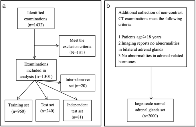
Flowchart of Data Collection and Grouping for (a) Adrenal Gland Segmentation Development Dataset from 1432 Non-contrast CT Imagings (2018–2020) and (b) Large-Scale Normal Adrenal Glands Dataset of 2000 Subjects for Age-Related Volume Analysis
Image acquisition and preprocessing
Non-contrast CT imaging of the adrenal gland was performed for all subjects using four CT scanners: Siemens Healthineers, Somatom Force, UIH uCT, and Philips Spectral 256-slice CT. The acquisition parameters were as follows: tube voltage of 120 kV, tube current of 104–267 mAs, matrix size of 512 × 512, and reconstructed slice thickness and interval of 1 mm. The 1301 CT examinations in the development dataset were distributed across the scanners as follows: Siemens Healthineers (n = 456), Somatom Force (n = 314), UIH uCT (n = 310), and Philips Spectral 256-slice CT (n = 221). To minimize the impact of varying scanning parameters, all CT images were resampled to an isotropic voxel resolution of 1 mm × 1 mm × 1 mm to ensure consistent spatial scaling. Additionally, window width and level standardization were applied using a window width of 400 Hounsfield Units (HU) and a window level of 40 HU to optimize image contrast and highlight the structural details of the target region. The grayscale values of the CT images were normalized to a range of 0–1 or constrained within − 1000 to 1000 HU to reduce the influence of different scanning devices.
Adrenal gland segmentation workflow and deep learning model construction
Manual annotation and segmentation workflow
Uploaded all 1301 CT images to the uAI Research Portal (Shanghai United Imaging Intelligence Co. Ltd, version 20231215) [29] in DICOM format. Two radiologists (with 6 and 7 years of experience, respectively) manually segmented the adrenal parenchyma of each side in axial images. Subsequently, the segmentations were reviewed by a senior radiologist (with 20 years of experience in diagnostic imaging). The results deemed accurate by the senior radiologist were established as the ground truth for neural network training and for the objective quantification of segmentation performance.
The adrenal gland segmentation workflow is illustrated in Fig. 2. The process begins with input 2D CT images and 3D CT volumes, annotated with a Region of Interest (ROI) and a Volume of Interest (VOI), respectively. The ROI is a 2D mask manually defined on a single axial slice to encompass the adrenal gland, while the VOI is 3D volumetric regions generated by stacking the 2D ROIs across all slices containing adrenal glands, providing volumetric context. Preprocessing involves cropping, resampling, normalization, and data augmentation. The preprocessed data were then fed into 2D and 3D nnU-Net models for training and testing, using 5-fold cross-validation to optimize performance, followed by best model selection and external testing. Post-processing included connected component analysis to refine segmentation by removing disconnected artifacts and calculating the adrenal gland volume.

Workflow of Adrenal Gland Segmentation Using nnU-Net on Non-contrast CT Images. This diagram shows adrenal gland segmentation using nnU-Net on non-contrast CT images. Inputs are 2D/3D CT images with ROI/VOI annotations (2D/3D masks). Preprocessing includes cropping, resampling, normalization, and data augmentation. Data are processed by 2D/3D nnU-Net with 5-fold cross-validation, followed by external testing. Post-processing refines segmentation and calculates volume
Deep learning segmentation model construction
We employed the nnU-Net framework, renowned for its robustness, self-configuring design and adaptability to various medical segmentation challenges, to train our segmentation model. A curated dataset of 1301 CT scans was utilized in the model’s training and testing. To compensate for the variability in scan quality and anatomical differences, CT images from Fuwai Central-China Cardiovascular Hospital featuring a diverse mix of male and female patients across different age groups were included.
This framework encouraged us to harness the effectiveness of this remarkable network as the backbone to refactor a new training pipeline according to our data, using the adrenal glands automatic segmentation approach. Recognizing the peculiar characteristics of medical image data, data augmentation techniques such as rotation, zoom, and adding noise were applied, but not the mirroring method, since the adrenal glands were specifically defined as left or right. A single nnU-Net model was trained to simultaneously segment both the left and right adrenal glands, treating them as two distinct classes in a multi-label segmentation task. Specifically, the ground truth annotations for each patient included two labels: one for the left adrenal gland and one for the right adrenal gland, enabling the model to generate two separate output segmentations per patient (one for each gland). We deployed a data augmentation strategy with five-fold cross-validation to mitigate the risk of over-fitting during prediction. All five models remained true to the standard methods outlined in nnU-Net for ensemble-based inference. However, for the final model used in testing and subsequent analysis, we selected the single model with the best average Dice Similarity Coefficient (DSC) across the five folds, rather than using an ensemble of the five models, as the single model achieved high performance (median DSC: 0.899–0.904) and met our requirements for clinical applicability. The five-fold cross-validation was subsequently adopted to streamline the hyperparameters. The optimal learning rate, batch size, and network depth were auto adjusted and optimized based on the input data. Each model underwent 1000 iterations. Besides, the optimal model was employed in an independent test set for model evaluation. The programming language of choice was Python 3.7.2 and it was executed on the PyTorch platform, utilizing two NVIDIA TITAN Xp graphic cards on a Linux operating system (Ubuntu 16.04 STL). Metrics such as the Dice Similarity Coefficient (DSC), Hausdorff Distance (HD), and Average Surface Distance (ASSD) between the automatically segmented and ground truth images were analyzed.
Inter-observer variability assessment
In light of the need for robust validation, we measured the performance of our segmentation model by gauging its consistency against the expertise of human radiologists. We had five board-certified radiologists provide annotations on a subset of 20 random cases, conducted in a blinded manner. Each radiologist independently outlined the adrenal glands on each provided CT image, after which we conducted a comparative analysis between the segmentations produced by the model and each of the radiologist’s annotations.
Statistical analysis
Statistical analyses were performed using MedCalc (version 19.8, MedCalc Software, Mariakerke, Belgium), R (version 4.1.3, Vienna, Austria), and Python (version 3.10.6). All statistical tests were two-sided, and P-values lower than 0.05 were considered statistically significant. For patient demographics comparison, continuous variables (e.g., age) were assessed for normality using the Shapiro-Wilk test; the Mann-Whitney U test was applied for non-normally distributed variables, and the chi-square test was used for categorical variables (e.g., sex). Model segmentation performance was evaluated using the Dice Similarity Coefficient (DSC), Hausdorff Distance (HD), and Average Surface Distance (ASSD), reported as medians with interquartile ranges (IQRs) due to non-normal distribution. Inter-observer agreement between radiologists and the model was compared using a two-sample t-test for DSC values. For age-related adrenal gland volume analysis, volumes were compared across age groups and sexes using the Kruskal-Wallis test (non-normally distributed data), followed by post-hoc Dunn’s test for pairwise comparisons.
Results
Patient and data set characteristics
The demographic characteristics of the training and test sets, independent test set, inter-observer set, and large-scale normal adrenal glands set are summarized in Table 1. Patient ages were recorded as integers in years, with medians and IQRs reported to one decimal place for consistency. There was no significant difference in age and sex between groups.
Segmentation results
Model performance on test and independent test sets
On the test set of 240 CT examinations in the five-fold cross-validation, the 3D nnU-Net median DSC scores are 0.899 and 0.904, the median HDs are 3.819 and 2.529, the median ASSDs are 0.075 and 0.067 in the left and right adrenal glands, respectively. The final independent test set included CT examinations of 81 cases, left and right adrenal glands 3D nnU-Net DSC scores are 0.900 and 0.896, the HDs are 6.612 and 5.361, the ASSDs are 0.091 and 0.077. The results indicated that there was a generally excellent agreement between machine and manual segmentation of adrenal glands. Table 2 summarizes the results in test set and independent test set.
Inter-observer variability and model consistency
Median inter-observer DSC ranged from 0.781 to 0.973 for 20 CT examinations. Median model-observer DSC ranged from 0.791 to 0.909 for all scans. The inter-observer DSC was not different from the model-observer DSC (P = 0.541). It was shown that the results of manual segmentation by five radiologists achieved a high degree of agreement. The results in the inter-observer set were summarized in Table 3. Annotations of an example case are shown in Fig. 3.

Comparison of Left Adrenal Gland Segmentation by Five Radiologists and nnU-Net in One Patient. Radiologists annotated the gland on a non-contrast CT image. Median inter-observer DSC was 0.781–0.973, and model-observer DSC was 0.791–0.909 (P = 0.541), showing high agreement
Age-related changes in adrenal gland volume
Understanding age-related changes in adrenal gland volume is crucial for clinical diagnosis and management, as these changes may reflect physiological adaptations or indicate underlying conditions such as adrenal hyperplasia or atrophy, which are associated with hormonal imbalances and metabolic disorders. In this study, we analyzed the bilateral adrenal gland volumes in a large-scale dataset of 2000 normal subjects (1328 males and 672 females) with no clinically indicated abnormalities, using the trained nnU-Net model for segmentation (Fig. 1b). The model used was the final nnU-Net model selected from the five-fold cross-validation, which achieved the best average DSC on the training and test sets. Subjects were grouped by 20-year age intervals, and volumes were calculated separately for males and females (Table 4). As shown in Fig. 4, adrenal gland volume generally increases with age up to the 40–60 age range, peaking at approximately 5.2 cm³ for males and 4.8 cm³ for females, before gradually decreasing in older age groups (60–80 years) to around 4.5 cm³ for males and 4.0 cm³ for females. Males consistently exhibited larger adrenal gland volumes than females across all age groups, with an average difference of 0.4–0.7 cm³ (Kruskal-Wallis test, P < 0.001; post-hoc Dunn’s test, P < 0.05 for male-female comparisons). These findings suggest that age and sex are significant factors influencing adrenal gland volume in healthy individuals.

Age-Related Changes in Bilateral Adrenal Gland Volume in Normal Subjects by Sex (Male: n = 1328; Female: n = 672). This graph shows age-related changes in bilateral adrenal gland volume in 2000 normal subjects, segmented by nnU-Net. Subjects were grouped by 20-year age intervals, with males (n = 1328, upper curve) and females (n = 672, lower curve) analyzed separately. Volume increases then decreases with age, with males having larger volumes
Discussion
Adrenal glands are small retroperitoneal organs that are difficult to segment accurately. The primary aim of this study is to develop a non-contrast CT-based deep learning algorithm for automated segmentation of adrenal glands. Our model achieved an exemplary performance, with median DSC scores of 0.899 and 0.904 for the left and right adrenal glands respectively in the test set, and 0.900 and 0.896 respectively in the independent test set.
Despite the successful segmentation of abdominal organs such as the liver and kidney in recent studies, only a few studies have performed on adrenal automated segmentation methods [30,31,32]. For instance, Robinson-Weiss C et al. [33] utilized a machine learning automatic segmentation model of the adrenal gland to achieve recognition of normal adrenal glands and adrenal glands with masses on enhanced CT images. The median DSCs for this segmentation model were 0.830 and 0.810. Moreover, Kim TM et al. [31] used an automated segmentation model in 308 abdominal CT scans to classify adrenal hyperplasia, the median DSCs of this segmentation model achieved 0.701. In our work, we have constructed a deep-learning algorithm for adrenal gland segmentation based on the 3D nnU-Net model. Notably, our model performs segmentation on non-contrast CT images, which offers a safer and more accessible alternative to contrast-enhanced CT by avoiding the risks associated with contrast agents, such as allergic reactions or renal impairment. However, the performance of our model on contrast-enhanced CT images remains untested, and future studies are needed to compare its effectiveness across different imaging modalities.
nnU-Net [32] is a type of deep learning model, specifically designed and optimized for medical image segmentation tasks. It is built upon the U-Net, but self-adaptive and more efficient. It includes various architectural modifications, cost function components and a standardized training scheme which are data-agnostic and enable the model to automatically adapt to the given task. In the context of CT adrenal gland segmentation, nnU-Net offers several advantages. First, it provides consistent outcomes since it eliminates the subjectivity associated with manual delineation. Second, it greatly improves efficiency, being able to process images faster than a human expert and thus could handle a larger amount of data.
The application of 3D nnU-Net to adrenal gland segmentation in CT not only accelerates the process and increases its objectivity, but also ensures excellent and reliable results for further research. This is crucial as accurate adrenal gland sketching lays the groundwork for subsequent analysis or disease detection. Therefore, the benefits of utilizing nnU-Net extend beyond operational efficiency and open up new avenues for adrenal gland-related clinical studies.
Our analysis of age-related adrenal gland volume changes provides valuable insights into normal adrenal morphology across the lifespan. The observed trend of volume increase until middle age followed by a decline in older age may reflect age-related physiological changes, such as increased adrenal activity during adulthood and potential atrophy in later years. The consistent volume difference between males and females highlights the influence of sex on adrenal gland morphology, which may be linked to hormonal differences. These findings can serve as a reference for identifying abnormal adrenal volumes in clinical settings, aiding in the diagnosis of conditions like adrenal hyperplasia or atrophy, and guiding further research into age- and sex-related adrenal disorders.
Despite these encouraging findings, our study does have some limitations. Primarily, it was a single-center study, the results of the segmentation performance of our model in a general population are not definitive yet. Additionally, our model was not tested on publicly available datasets, which limits the reproducibility of our results in external validation settings. In future work, we will include images from other centers, set up additional external test sets, and evaluate our model on publicly available datasets to improve its generalizability and robustness. Furthermore, excluding cases where adrenal glands were indistinguishable from surrounding tissues may have reduced the task’s complexity, potentially leading to higher DSC scores and limiting performance in challenging clinical scenarios. Also, the dataset’s male-to-female ratio of 2:1 (Table 1) may affect model performance due to anatomical differences, but gender-specific performance was not analyzed in this study.
Future studies will address these limitations by including images from multiple centers and publicly available datasets to enhance generalizability, incorporating challenging cases to test the model in complex clinical scenarios, and analyzing gender-specific performance to ensure robustness across diverse populations.
In conclusion, the established non-contrast CT-based deep learning algorithm can competently automate the segmentation of adrenal glands.
Data availability
The datasets used and analyzed during the current study are available from the corresponding author on reasonable request.
References
Navin PJ, Moynagh MR. Optimal and novel imaging of the adrenal glands. Curr Opin Endocrinol Diabetes Obes. 2022;29(3):253–62. https://doiorg.publicaciones.saludcastillayleon.es/10.1097/MED.0000000000000730.
Ctvrtlik F, Koranda P, Tichy T. Adrenal disease: a clinical update and overview of imaging. A review. Biomed Pap Med Fac Univ Palacky Olomouc Czech Repub. 2014;158(1):23–34. https://doiorg.publicaciones.saludcastillayleon.es/10.5507/bp.2014.010.
Donnellan WL. Surgical anatomy of adrenal glands. Ann Surg. 1961;154(Suppl 6):298–305. https://doiorg.publicaciones.saludcastillayleon.es/10.1097/00000658-196112000-00040.
Mete O, Erickson LA, Juhlin CC, et al. Overview of the 2022 WHO classification of adrenal cortical tumors. Endocr Pathol. 2022;33(1):155–96. https://doiorg.publicaciones.saludcastillayleon.es/10.1007/s12022-022-09710-8.
Mohan DR, Lerario AM. Closing the loop on adrenal health, dysfunction, and disease. Sci Transl Med. 2023;15(701):eadh4450. https://doiorg.publicaciones.saludcastillayleon.es/10.1126/scitranslmed.adh4450.
Ebbehoj A, Li D, Kaur RJ, et al. Epidemiology of adrenal tumours in olmsted County, Minnesota, USA: a population-based cohort study. Lancet Diabetes Endocrinol. 2020;8(11):894–902. https://doiorg.publicaciones.saludcastillayleon.es/10.1016/S2213-8587(20)30314-4.
Park SS, Kim JH. Recent updates on the management of adrenal incidentalomas. Endocrinol Metab (Seoul). 2023;38(4):373–80. https://doiorg.publicaciones.saludcastillayleon.es/10.3803/EnM.2023.1779.
Bancos I. Adrenal incidentalomas: insights into prevalence. Ann Intern Med. 2022;175(10):1481–2. https://doiorg.publicaciones.saludcastillayleon.es/10.7326/M22-2600.
Kebebew E, Adrenal Incidentaloma. N Engl J Med. 2021;384(16):1542–51. https://doiorg.publicaciones.saludcastillayleon.es/10.1056/NEJMcp2031112.
Fassnacht M, Tsagarakis S, Terzolo M, et al. European society of endocrinology clinical practice guidelines on the management of adrenal incidentalomas, in collaboration with the European network for the study of adrenal tumors. Eur J Endocrinol. 2023;189(1):G1–42. https://doiorg.publicaciones.saludcastillayleon.es/10.1093/ejendo/lvad066.
Libianto R, Yang J, Fuller PJ. Adrenal disease: an update. Aust J Gen Pract. 2021;50(1–2):9–14. https://doiorg.publicaciones.saludcastillayleon.es/10.31128/AJGP-09-20-5619.
Sam D, Kline GA, So B, et al. Unilateral disease is common in patients with primary aldosteronism without adrenal nodules. Can J Cardiol. 2020;37(2):269–75. https://doiorg.publicaciones.saludcastillayleon.es/10.1016/j.cjca.2020.05.013.
Arnaldi G, Boscaro M, Adrenal incidentaloma. Best Pract Res Clin Endocrinol Metab. 2012;26(4):405–19. https://doiorg.publicaciones.saludcastillayleon.es/10.1016/j.beem.2011.12.006.
Li J, Li H, Zhang Y, et al. MCNet: A multi-level context-aware network for the segmentation of adrenal gland in CT images. Neural Netw. 2023;170:136–48. https://doiorg.publicaciones.saludcastillayleon.es/10.1016/j.neunet.2023.11.028.
Charoensri S, Auchus RJ. A contemporary approach to the diagnosis and management of adrenal insufficiency. Endocrinol Metab (Seoul). 2024;39(1):73–82. https://doiorg.publicaciones.saludcastillayleon.es/10.3803/EnM.2024.1894.
Xiao DX, Zhong JP, Peng JD, et al. Machine learning for differentiation of lipid-poor adrenal adenoma and subclinical pheochromocytoma based on multiphase CT imaging radiomics. BMC Med Imaging. 2023;23(1):159. Published 2023 Oct 16. https://doiorg.publicaciones.saludcastillayleon.es/10.1186/s12880-023-01106-2
Loper MR, Makary MS. Evolving and novel applications of artificial intelligence in abdominal imaging. Tomography. 2024;10(11):1814–31. https://doiorg.publicaciones.saludcastillayleon.es/10.3390/tomography10110133
Föllmer B, Williams MC, Dey D, et al. Roadmap on the use of artificial intelligence for imaging of vulnerable atherosclerotic plaque in coronary arteries. Nat Rev Cardiol. 2023;21(1):51–64. https://doiorg.publicaciones.saludcastillayleon.es/10.1038/s41569-023-00900-3.
Nurmohamed NS, Bom MJ, Jukema RA, et al. AI-Guided quantitative plaque staging predicts Long-Term cardiovascular outcomes in patients at risk for atherosclerotic CVD. JACC Cardiovasc Imaging. 2024;17(3):269–80. https://doiorg.publicaciones.saludcastillayleon.es/10.1016/j.jcmg.2023.05.020.
Dey D, Arnaout R, Antani S, et al. Proceedings of the NHLBI Workshop on Artificial Intelligence in Cardiovascular Imaging: Translation to Patient Care. JACC Cardiovasc Imaging. 2023;16(9):1209–1223. https://doiorg.publicaciones.saludcastillayleon.es/10.1016/j.jcmg.2023.05.012
Deng Y, Wang H, He L. CT radiomics to differentiate between Wilms tumor and clear cell sarcoma of the kidney in children. BMC Med Imaging. 2024;24(1):13. https://doiorg.publicaciones.saludcastillayleon.es/10.1186/s12880-023-01184-2
Yin X, Liao H, Yun H, et al. Artificial intelligence-based prediction of clinical outcome in immunotherapy and targeted therapy of lung cancer. Semin Cancer Biol. 2022;86(Pt 2):146–59. https://doiorg.publicaciones.saludcastillayleon.es/10.1016/j.semcancer.2022.08.002.
Chen M, Copley SJ, Viola P, Lu H, Aboagye EO. Radiomics and artificial intelligence for precision medicine in lung cancer treatment. Semin Cancer Biol. 2023;93:97–113. https://doiorg.publicaciones.saludcastillayleon.es/10.1016/j.semcancer.2023.05.004.
Jiang J, Chao WL, Culp S, et al. Artificial intelligence in the diagnosis and treatment of pancreatic cystic lesions and adenocarcinoma. Cancers (Basel). 2023;15(9). https://doiorg.publicaciones.saludcastillayleon.es/10.3390/cancers15092410.
Khan RA, Luo Y, Wu FX, RMS-UNet. Residual multi-scale UNet for liver and lesion segmentation. Artif Intell Med. 2022;124:102231. https://doiorg.publicaciones.saludcastillayleon.es/10.1016/j.artmed.2021.102231.
Yu Y, He Z, Ouyang J, et al. Magnetic resonance imaging radiomics predicts preoperative axillary lymph node metastasis to support surgical decisions and is associated with tumor microenvironment in invasive breast cancer: A machine learning, multicenter study. EBioMedicine. 2021;69:103460. https://doiorg.publicaciones.saludcastillayleon.es/10.1016/j.ebiom.2021.103460.
Wang S, Summers RM. Machine learning and radiology. Med Image Anal. 2012;16(5):933–51. https://doiorg.publicaciones.saludcastillayleon.es/10.1016/j.media.2012.02.005.
Koyuncu H, Ceylan R, Erdogan H, et al. A novel pipeline for adrenal tumour segmentation. Comput Methods Programs Biomed. 2018;159:77–86. https://doiorg.publicaciones.saludcastillayleon.es/10.1016/j.cmpb.2018.01.032.
Wu J, Xia Y, Wang X, et al. uRP: an integrated research platform for one-stop analysis of medical images. Front Radiol. 2023;3:1153784. https://doiorg.publicaciones.saludcastillayleon.es/10.3389/fradi.2023.1153784
Saiprasad G, Chang CI, Safdar N, et al. Adrenal gland abnormality detection using random forest classification. J Digit Imaging. 2013;26(5):891–7. https://doiorg.publicaciones.saludcastillayleon.es/10.1007/s10278-012-9554-7.
Kim TM, Choi SJ, Ko JY, et al. Fully automatic volume measurement of the adrenal gland on CT using deep learning to classify adrenal hyperplasia. Eur Radiol. 2023;33(6):4292–302. https://doiorg.publicaciones.saludcastillayleon.es/10.1007/s00330-022-09347-5.
Isensee F, Jaeger PF, Kohl SAA, Petersen J, Maier-Hein KH. nnU-Net: a self-configuring method for deep learning-based biomedical image segmentation. Nat Methods. 2021;18(2):203–11. https://doiorg.publicaciones.saludcastillayleon.es/10.1038/s41592-020-01008-z.
Robinson-Weiss C, Patel J, Bizzo BC, et al. Machine learning for adrenal gland segmentation and classification of normal and adrenal masses at CT. Radiology. 2023;306(2):e220101. https://doiorg.publicaciones.saludcastillayleon.es/10.1148/radiol.220101.
Acknowledgements
Not applicable.
Funding
This work was supported by Henan Cardiovascular Disease Center (Central China Subcenter of National Center for Cardiovascular Diseases) (Grant No. 2023-FZX17), China, and Henan Provincial Key Laboratory of Cardiology Medical Imaging (Grant No. 2021-44-16), China.
Author information
Authors and Affiliations
Contributions
F.M., T.Z., X.K. and Y.P. contributed to the conceptualization; F.M., T.Z., M.X., J.C. and F.L. contributed to the acquisition of data; F.L., F.M., T.Z. and Y.X. contributed to the analysis of data; F.M. contributed to the writing of the original draft and visualization. Y.G. and Y.X. edited the manuscript. All authors reviewed and approved the final version of the manuscript.
Corresponding author
Ethics declarations
Ethics approval and consent to participate
This study was approved by the Ethics Committee of Fuwai Central-China Cardiovascular Hospital (Approval No. 2025.21). Given the retrospective nature of the study and the use of fully anonymized patient data, the requirement for informed consent was waived by the ethics committee. All procedures were conducted in accordance with the ethical standards outlined in the Declaration of Helsinki and its later amendments.
Consent for publication
Not applicable.
Competing interests
The authors declare no competing interests.
Additional information
Publisher’s note
Springer Nature remains neutral with regard to jurisdictional claims in published maps and institutional affiliations.
Electronic supplementary material
Below is the link to the electronic supplementary material.
Rights and permissions
Open Access This article is licensed under a Creative Commons Attribution-NonCommercial-NoDerivatives 4.0 International License, which permits any non-commercial use, sharing, distribution and reproduction in any medium or format, as long as you give appropriate credit to the original author(s) and the source, provide a link to the Creative Commons licence, and indicate if you modified the licensed material. You do not have permission under this licence to share adapted material derived from this article or parts of it. The images or other third party material in this article are included in the article’s Creative Commons licence, unless indicated otherwise in a credit line to the material. If material is not included in the article’s Creative Commons licence and your intended use is not permitted by statutory regulation or exceeds the permitted use, you will need to obtain permission directly from the copyright holder. To view a copy of this licence, visit http://creativecommons.org/licenses/by-nc-nd/4.0/.
About this article

Cite this article
Meng, F., Zhang, T., Pan, Y. et al. A deep learning algorithm for automated adrenal gland segmentation on non-contrast CT images. BMC Med Imaging 25, 142 (2025). https://doiorg.publicaciones.saludcastillayleon.es/10.1186/s12880-025-01682-5
Received:
Accepted:
Published:
DOI: https://doiorg.publicaciones.saludcastillayleon.es/10.1186/s12880-025-01682-5