- Research
- Open access
- Published:
Intelligent detection and grading diagnosis of fresh rib fractures based on deep learning
BMC Medical Imaging volume 25, Article number: 98 (2025)
Abstract
Background
Accurate detection and grading of fresh rib fractures are crucial for patient management but remain challenging due to the complexity of rib structures on CT images.
Methods
Chest CT images from 383 patients with rib fractures were retrospectively analyzed. The dataset was divided into a training set (n = 306) and an internal testing set (n = 77). An external testing set of 50 patients from the public RibFrac dataset was included. Fractures were classified into severe and non-severe categories. A modified YOLO-based deep learning model was developed for detection and grading. Performance was compared with thoracic surgeons using precision, recall, mAP50, and F1 score.
Results
The deep learning model showed excellent performance in diagnosing fresh rib fractures. For all fractures types in internal test set, the precision, recall, mAP50, and F1 score were 0.963, 0.934, 0.972, and 0.948, respectively. The model outperformed thoracic surgeons of varying experience levels (all p < 0.01).
Conclusion
The proposed deep learning model can automatically detect and grade fresh rib fractures with accuracy comparable to that of physicians. This model helps improve diagnostic accuracy, reduce physician workload, save medical resources, and strengthen health care in resource-limited areas.
Clinical trial number
Not applicable.
Introduction
Rib fractures are the most common injuries in patients with blunt chest trauma [1, 2] and may be accompanied by severe complications such as pulmonary contusion, pneumonia, and hemopneumothorax [3, 4]. The number and displacement of rib fractures, as well as the presence of associated organ injuries, are correlated with mortality [5, 6]. Therefore, accurately identifying the number and severity of rib fractures is crucial.
With the increased use of chest multidetector computed tomography scans, the detection rate of rib fractures has significantly improved [7]. Compared to X-ray, CT offers greater contrast and resolution, providing a comprehensive view of the fracture site and enabling the detection of minor lesions and the assessment of other complications. However, the diversity and complexity of rib fracture shapes make it challenging to identify all fractures across hundreds of thin-slice CT images, often leading to missed diagnoses. The literature reports that 20.7% of initial chest CT scan reports misidentify the number or location of fractured ribs [8], and even after rib reconstruction, 20.9% of rib fractures are missed [9].
Recently, deep learning algorithms have been applied in the field of medical image processing [10], including image registration [11, 12], detection [13,14,15], segmentation [16,17,18] and disease prognosis [19, 20]. Notably, studies focusing on Alzheimer’s disease [21] and brain tumor classification [22], have highlighted the potential of next-generation convolutional architectures to enhance diagnostic accuracy and efficiency. Building upon these advancements, our study applies a deep learning approach to the detection and grading of fresh rib fractures. This approach not only aims to assist clinicians in identifying fractures more accurately but also seeks to alleviate their workload and ultimately improve patient outcomes. Several studies have reported promising results in rib fractures detection [23,24,25,26]. Azuma et al. [27] developed and validated a convolutional neural network (CNN) model for detecting rib fractures, showing that the CNN model could enhance the diagnostic ability of radiologists for any type of rib fracture. Zhou et al. [26] included clinical information in their CNN model, further improving diagnostic efficiency and reducing diagnosis time. However, the importance of fractures varies with their type. Chronic rib fractures usually do not require clinical intervention and do not affect treatment decisions or patient prognosis. In contrast, fresh rib fractures often exhibit severe displacement and may be accompanied by organ injuries, especially severely displaced and comminuted rib fractures, which are often accompanied by fatal complications such as severe pulmonary infection, persistent hemothorax, and massive hemoptysis [28]. Moreover, rapid and accurate detection of the severity of fresh rib fractures helps to implement necessary treatment measures, thereby improving patient prognosis. Therefore, accurately identifying the location and severity of fresh rib fractures is particularly important. To the best of our knowledge, there is a scarcity of studies focusing on the graded diagnosis of fresh rib fractures, with an emphasis on those that are severely displaced.
In our study, we developed a deep learning-based model for the intelligent detection of fresh rib fractures and graded diagnoses based on the severity of rib displacement, categorizing fractures into severe and non-severe. We conducted a comprehensive analysis of different subgroups of fracture severity and compared the model’s performance with that of experienced thoracic surgeons.
Methods
Data sets and classification criteria
A retrospective collection of initial chest CT scans of patients diagnosed with rib fractures from January 2021 to April 2023 was conducted at the First Affiliated Hospital of Army Medical University (Hospital A) and Chongqing Dianjiang People’s Hospital (Hospital B). This study obtained ethical approval from the Ethics Committee of the First Affiliated Hospital of Army Medical University (ID: KY2023062) and Chongqing Dianjiang People’s Hospital (ID: DYLL-LW-2023-03). All patient data were de-identified prior to analysis, and this retrospective study was conducted in accordance with the Declaration of Helsinki. The inclusion criteria were patients aged ≥ 18 years with a history of trauma. Exclusion criteria included: (1) old or healed fractures, (2) artifacts, (3) bone tumors or bone destruction, (4) postoperative patients.
A total of 383 patients from the two centers were randomly divided into a training set (n = 306) and an internal testing set (n = 77). A rigorous randomization method was employed to minimize bias introduced by data partitioning and ensure the reliability and scientific validity of the study results. The external testing dataset comprised 50 CT scans from the public RibFrac dataset [29], and radiologists re-annotated the CT scans of these 50 cases. The specific screening criteria are shown in Fig. 1.
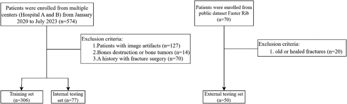
Flow chart of patient selection
In this study, fractures were classified into non-severe and severe based on the severity. Non-severe fractures were defined as: (1) fissure fractures with bone defects between the ends of the cortical bone without displacement, (2) cortical bone distortion or density changes, (3) fractures with angulation or displacement less than the rib diameter. Severe fractures were defined as: (1) fractures with displacement greater than the rib diameter, (2) fractures with observed bone fragments or comminuted fractures (Fig. 2).

Examples of two different types of rib fractures. The top row shows non-severe fractures, while the bottom row shows severe fractures. (a) Mild fracture with cortical distortion, where the outer layer of the rib is slightly disrupted but remains mostly intact. (b) Mild fracture with misalignment or displacement, but the displacement distance is less than the rib’s diameter. (c) Severe comminuted fracture, with the rib broken into multiple fragments. (d) Severe fracture with significant displacement, where the displacement distance exceeds the rib’s diameter
CT acquisition
Patients were positioned supine, with their arms raised above their heads (for those unable to do so due to shoulder or upper limb injuries, arms were placed naturally at their sides). The scanning range extended from the thoracic inlet to the lung bases or the entire ribcage. The scan was performed during a single breath-hold following inhalation. Two different CT scanners were utilized for the examinations: Philips Brilliance 16 (Philips Medical Systems) and SOMATOM Definition (Siemens Healthineers). The tube voltage was set to 120 kV, with automatic modulation of the tube current. The slice interval ranged from 1 to 5 mm, and the slice thickness varied between 1 and 5 mm.
Image annotation
The CT slices in the training set were independently annotated by a thoracic surgeon with 5 years of experience and a radiologist with 5 years of experience. These two physicians used Makesense (https://www.makesense.ai/) to mark rib fractures with rectangular boxes and used different colors to indicate severity (Supplementary Fig. 1). In cases of uncertainty regarding fracture type, the two physicians discussed until a consensus was reached, or a third physician was involved to reach a final agreement. Annotations were made without access to any clinical information of the patients, and these annotations were used as the ground truth for training the automated detection algorithm.
The internal and external test datasets were annotated independently by another three thoracic surgeons -a junior, a middle grade, and a senior, and these annotations were subsequently compared with the model’s predictions.
Model architecture
The model architecture: You-Only-Look-Once (YOLO) version 8 [30] is a real-time object detection algorithm that can detect and locate multiple objects in images or videos with relatively fast speed. While maintaining high detection accuracy, it significantly improves reasoning speed and is suitable for various computing devices, including mobile devices and embedded systems. The model architecture consists of three main components: the Backbone, the Neck, and the Head networks.
Although YOLOv8 has achieved notable success in fields such as autonomous driving and industrial inspection, it still exhibits significant shortcomings in the detection of small targets in medical imaging. To enhance YOLOv8’s capability in detecting small targets, we propose improvements by incorporating high-resolution feature maps and introducing a more sophisticated Feature Pyramid Network (FPN). The specific innovations are as follows:
1. Design of the C2f_EMA Module in YOLOv8: A C2f_EMA module was designed in YOLOv8, which incorporates the EMA (Efficient Multi-scale Attention) attention mechanism. This modular design retains the original feature extraction capability of the C2f module while enhancing feature representation through the attention mechanism.
2. Integration of the Attention Mechanism: By embedding the EMA attention mechanism into the C2f module of YOLOv8, the study achieves dynamic adjustment of channel and spatial dimension weights during the feature extraction and fusion stages, thereby improving the model’s discriminative ability.
3. Improved Feature Pyramid Network: The study enhances the FPN-PAN structure of YOLOv8 by introducing the new C2f_EMA module. This improvement facilitates the construction of a more efficient feature pyramid network, leading to enhanced model performance.
Figure 3 illustrates the process of detecting rib fractures using the improved YOLOv8 model architecture with the C2f_EMA module and enhanced Feature Pyramid Network, showcasing how feature maps are processed and the final detection result is achieved. These innovations collectively aim to address the limitations of YOLOv8 in small target detection, particularly in the context of medical imaging, by improving feature extraction, fusion, and multi-scale representation capabilities. These innovations collectively aim to address the limitations of YOLOv8 in small target detection, particularly in the context of medical imaging, by improving feature extraction, fusion, and multi-scale representation capabilities.

Rib fracture detection in CT images using a YOLO-based model with Exponential Moving Average (EMA). The input CT images are resized to 640px × 640px and processed through the model to generate feature maps. These maps are used to produce a class probability map and a bounding map, which are then passed through Non-Maximum Suppression (NMS) to yield the final detection of the fracture location
As shown in Fig. 4, the input image is first passed through the Backbone to extract multi-scale features. The Backbone network (e.g., CSPDarknet53) generates multi-scale feature maps through convolutional layers and downsampling operations [31, 32] capturing semantic information from low-level to high-level layers, thereby enhancing the feature extraction capability for small targets. To improve feature representation, we introduce the C2f_EMA module, which combines the C2f structure with the EMA (Efficient Multi-scale Attention) mechanism to dynamically adjust channel and spatial weights, enhancing the focus on important features.

The architecture of a YOLO-based model incorporating Exponential Moving Average (EMA). The backbone extracts features using CBS and C2f_EMA modules, followed by spatial pyramid pooling (SPPf) and feature fusion in the neck. The detection head generates outputs across multiple scales for object detection
Subsequently, the multi-scale features are fed into the Neck module for feature fusion. The Neck adopts the FPN (Feature Pyramid Network) and PAN (Path Aggregation Network) structures, aggregating features through top-down and bottom-up paths to enhance multi-scale object detection capabilities. We also integrate the C2f_EMA module into the Neck, further optimizing the feature pyramid network and improving feature fusion performance.
Through the collaborative work of the Backbone and Neck, combined with the C2f_EMA module, the model can more efficiently extract and fuse multi-scale features, providing richer feature representations for the detection head (Head). This significantly improves the accuracy and robustness of small target detection, particularly excelling in complex scenarios such as medical imaging.
\(\:x\in\:{\mathbb{R}}^{H\times\:W\times\:3}\), where Fb represents multi-scale features, Fn is the fused feature map, and Y is the output of the detection head.
Model evaluation
To objectively assess the model’s performance, four evaluation metrics were calculated: mean average precision (mAP), precision, recall, and F1 score. For localization accuracy, mAP was used, which is a standard metric in artificial intelligence. The areas under the precision-recall curve (AUPRC), with values ranging between 0 and 1. To calculate mAP, intersection of union (IoU) is used, measuring the overlap between predictions and ground truth. We defined the IoU threshold as 0.5 to classify the prediction box as a true positive or false positive. An IoU of 0.5 was chosen because, in clinical CT examinations, such a prediction box can already prompt clinicians (Supplementary Fig. 2). Precision is defined as the number of correctly predicted slices divided by the total number of predicted slices, while recall is defined as the number of samples correctly predicted as positive by the model divided by the total number of positive samples. The F1 score is the harmonic mean of precision and recall, providing a balance between the two metrics. It is particularly useful when the class distribution is imbalanced.
Statistical analysis
This study employed SPSS (version 26.0, IBM, NY for statistical analysis, with a two-tailed p-value threshold of < 0.05. Normality of continuous variables was assessed using the Kolmogorov-Smirnov test. Normally distributed data were presented as mean ± standard deviation, otherwise, M (P25-P75) were utilized. Categorical variables were expressed as frequencies and percentages.
For normally distributed data, two-sample t-tests were applied, and non-normally distributed data were analyzed using the Mann-Whitney U test. Categorical variables were compared using Chi-squared or Fisher’s exact test. The z-test was used to compare whether there was a statistical difference between the system and the thoracic surgeons.
Results
Patient characteristics
This study included data from 383 patients with rib fractures from two centers, comprising a total of 4673 annotations. Among these annotations, 3,043 were for non-severe fractures and 1,630 for severe fractures. Of the 383 patients, 130 were from Hospital A and 253 from Hospital B. We randomly selected 306 patients for the training set and 77 patients for the internal testing set. Our results showed no statistically significant differences in gender (P = 0.256) and age (P = 0.766) between the training and testing sets (Table 1). Additionally, there were no significant differences in age (P = 0.565) between patients from the two centers, and hospital A had a significantly higher proportion of male patients compared to hospital B (P < 0.01) as detailed in Supplementary Table 1.
The training set consisted of 306 patients, with a total of 3,646 annotations, including 2,455 non-severe fractures and 1,191 severe fractures. The internal testing set included 77 patients, with a total of 1,027 annotations, comprising 685 non-severe and 342 severe fractures. Additionally, we incorporated 50 patients from the public RibFrac dataset, with a total of 821 annotations, including 660 non-severe fractures and 161 severe fractures.
Performance of the models
In the internal test set, the overall precision for all fracture types was 0.963, recall was 0.934, mAP50 was 0.972, and the F1 score was 0.948. For external test dataset, precision was 0.880, recall was 0.796, mAP50 was 0.857, and F1 score was 0.836 (Table 2).
We visualized the model’s performance using the precision-recall (PR) curve. Figure 5a illustrates the PR curves for the internal test set, where the AUPRC values are 0.964 for non-severe fractures, 0.981 for severe fractures, and 0.972 for all classes combined. Figure 5b shows the PR curves for the external test set, with AUPRC values of 0.843 for Non-severe fractures, 0.872 for Severe fractures, and 0.857 for all classes combined. These results demonstrate that our model exhibits excellent performance on the external test set, indicating good generalizability. Figure 6 presents examples of rib fractures successfully identified by our model. Additionally, a confusion matrix has been created to illustrate the model’s performance on the internal and external testing set (Supplementary Fig. 3).

Precision-Recall curves depicting the model’s diagnostic performance for fracture identification. (a) Internal test set results: The model demonstrates high Average Precision (AP) scores, with an AP of 0.964 for non-severe fractures and 0.981 for severe fractures, culminating in a combined AP of 0.972 for all classes. (b) External test set results: The model maintains robust performance with APs of 0.843 for non-severe fractures, 0.872 for severe fractures, and an aggregate AP of 0.857 across all classes

Overview of fracture cases identified by the model, encompassing non-severe and severe fracture recognition cases. a and b: Non-severe fractures identified by the model, Fig. 6c and d: Severe fractures identified by the model
To further demonstrate the effectiveness of the model, we performed comparative experiments with the YOLOv8x and EfficientVit-M3 models, respectively, for the two different datasets and for non-severe fractures and severe fractures. The experimental results show that the F1-score of the proposed model on the internal verification set is higher than other models, and the accuracy and recall rate of the proposed model on the external verification set are 5% higher than other models, which further indicates the effectiveness of the model in detecting small targets (Table 3).
Comparison of the performance between our model and thoracic surgeons
Table 4, and Fig. 7 present a comparison of precision, recall, and F1 score between three thoracic surgeons of varying experience levels and our model. For non-severe fractures, the F1 score ranged from 0.660 to 0.862 across surgeons, with senior surgeon achieving the highest score. For severe fractures, senior surgeon again performed the best with an F1 score of 0.880, while junior surgeon had the lowest at 0.648. In overall performance, senior surgeon led with an F1 score of 0.869, while our model outperformed all surgeons in both precision and recall, demonstrating its superior capability in rib fracture detection. These results emphasize the model’s potential in improving detection accuracy compared to experienced clinicians.

Performance comparison between our model and doctors. (a): doctor’s performance on the precision-recall curve. (b): The Statistical results of precision, recall and F1 score on internal test dataset. mAP: mean average precision, JT: Junior Thoracic surgeon, MT: Middle grade Thoracic surgeon, SR: Senior Thoracic surgeon, Model: Deep learning system for fresh rib fracture detection and grading model
Discussion
Chest CT scans are often the preferred imaging modality for patients with chest trauma because they can identify many injuries that may be missed by chest X-rays, including pulmonary contusions, hemothorax, pneumothorax, and rib fractures. Moreover, rib fractures are considered indicators of severe trauma [33]. Traditional detection methods require physicians to meticulously evaluate the entire CT scan, which is a time-consuming and error-prone process, particularly for less experienced radiologists or thoracic surgeons. Additionally, the number and displacement of rib fractures are related to the follow-up treatment plan [34, 35]. Therefore, thoracic surgeons should prioritize different types of rib fractures. Chronic rib fractures, characterized by the presence of a mature callus or an invisible fracture line, do not require clinical intervention and do not affect treatment decisions or patient prognosis, making their detection unnecessary. In contrast, timely identification and localization of fresh rib fractures are crucial, especially in emergency settings, as severely displaced rib fractures may be associated with internal organ injuries, significantly impacting patient outcomes [36].
To address this issue, we developed a deep learning-based intelligent diagnostic model for the classification and detection of fresh rib fractures, which was validated on both internal multicenter datasets and external public datasets. The model outperformed experienced thoracic surgeons and demonstrated exceptional performance.
Recently, Yao et al. [37] developed a deep learning-based rib fracture detection system that achieved high-performance detection and diagnosis of rib fractures on chest CT images, significantly reducing physician workload and minimizing misdiagnoses. However, this study only addressed the binary classification of fracture presence. Zhou et al. [38] developed a convolutional neural network (CNN) model that classified fractures into fresh, healing, and old fractures but did not perform a graded diagnosis for precise fracture diagnosis. Zhou et al. [26] reported the use of clinical information in their CNN model, which similarly improved diagnostic efficiency and reduced diagnosis time. Xiong et al. [39] found that the performance of radiologists on night shifts was inferior to that on day shifts, and the use of a deep learning-based computer-aided diagnosis (CAD) system for rib fractures helped night shift radiologists achieve performance levels comparable to their daytime performance. Unlike previous studies, our research focused on fresh rib fractures, as their rapid localization and diagnosis are crucial components of intelligent diagnosis and treatment of acute chest trauma. Chronic rib fractures do not affect treatment decisions or patient outcomes, making the focus on fresh rib fractures more aligned with real clinical scenarios. Our model can also intelligently grade fractures based on their severity, aiding in treatment decisions and prognosis evaluation for posttraumatic rib fractures.
Our model’s performance is also comparable to that of physicians, making it suitable as the “first reader.” This approach can help improve diagnostic accuracy, reduce diagnosis time, and reduce the workload of physicians, additionally, it can aid in building medical resources in under resourced areas.
In our model improvements, we optimized the Backbone, Neck, and Head networks to enhance feature extraction, fusion, and detection capabilities. The Backbone, designed to extract deep features from medical images, alternates between CBS modules (Convolution, Batch Normalization, and SiLU activation) and C2f_EMA modules, which include convolution layers and parallel Bottleneck_EMA branches for richer feature representation. It concludes with the SPPF module, which combines multiple Maxpool and Concat operations to capture critical global information. In the Neck, we focused on refining and consolidating multi-scale features using an Upsample operation and the C2f_EMA module to merge and process features across different levels, improving detection accuracy and robustness. Finally, the Head network, composed of additional C2f_EMA modules, integrates and processes refined features before passing them to the Detect layer, where bounding boxes and class predictions are generated. This comprehensive multi-level integration allows our model to achieve high-precision detection in medical images.
Limitations of the study
Our study had several limitations. First, while the model can mark the fracture locations on CT images, it cannot provide the specific anatomical details, such as identifying which rib is fractured. Second, our sample size could be further expanded, and we plan to include more centers in future studies. Finally, prospective studies remain relatively scarce. We aim to collect more data and conduct prospective studies to further validate and optimize the model.
Conclusion
In summary, we developed a deep learning-based intelligent detection model for the detection and grading of fresh rib fractures. The model demonstrated high fracture detection rates and localization accuracy. It is suitable as a “first reader” to assist physicians in quickly and accurately determining the condition of patients with rib fractures, reducing diagnostic time, improving diagnostic accuracy, and enhancing physician efficiency. Additionally, the model can help strengthen medical resources in under resourced areas, including rural and township regions.
Data availability
The datasets used and analysed during the current study are available from the corresponding author on reasonable request.
Abbreviations
- AI:
-
Artificial intelligence
- CT:
-
Computed tomography
- CNN:
-
Convolutional neural network
- CAD:
-
Computer-aided diagnosis
- mAP:
-
mean average precision
- ROC-AUC:
-
Receiver operating characteristic - area under curve
- PR:
-
Precision-recall
- IoU:
-
Intersection over Union
- EMA:
-
Exponential moving average
- CBS:
-
Convolution, batch normalization, SiLU activation
- SPPF:
-
Spatial pyramid pooling in feature
- YOLO:
-
You only look once
- AUPRC:
-
Area under the precision-recall curve
References
Lin FC-F, Li R-Y, Tung Y-W, Jeng K-C, Tsai SC-S. Morbidity, mortality, associated injuries, and management of traumatic rib fractures. J Chin Med Assoc. 2016;79:329–34.
Marini CP, Petrone P, Soto-Sánchez A, García-Santos E, Stoller C, Verde J. Predictors of mortality in patients with rib fractures. Eur J Trauma Emerg Surg. 2021;47:1527–34.
Miller LA. Chest wall, lung, and pleural space trauma. Radiol Clin North Am 2006;44.
Talbot BS, Gange CP, Chaturvedi A, Klionsky N, Hobbs SK, Chaturvedi A. Traumatic rib injury: patterns, imaging pitfalls, complications, and treatment. Radiographics. 2017;37:628–51.
Brasel KJ, Moore EE, Albrecht RA, deMoya M, Schreiber M, Karmy-Jones R, Rowell S, Namias N, Cohen M, Shatz DV. Biffl, Western trauma association critical decisions in trauma: management of rib fractures. J Trauma Acute Care Surg. 2017;82:200–3.
Lee RB, Bass SM, Morris JA, MacKenzie EJ. Three or more rib fractures as an indicator for transfer to a level I trauma center: a population-based study. J Trauma. 1990;30:689–94.
Cherney AR, Richardson DM, Greenberg MR, Choo EK, McGregor AJ, Safdar B. Prevalence and clinical import of thoracic injury identified by chest computed tomography but not chest radiography in blunt trauma: multicenter prospective cohort study. Ann Emerg Med. 2016;68:133–4.
Cho SH, Sung YM, Kim MS. Missed rib fractures on evaluation of initial chest CT for trauma patients: pattern analysis and diagnostic value of coronal multiplanar reconstruction images with multidetector row CT. Br J Radiol. 2012;85:e845–50.
Urbaneja A, De Verbizier J, Formery A-S, Tobon-Gomez C, Nace L, Blum A, Gondim PA, Teixeira. Automatic rib cage unfolding with CT cylindrical projection reformat in polytraumatized patients for rib fracture detection and characterization: feasibility and clinical application. Eur J Radiol. 2019;110:121–7.
Bhatt C, Kumar I, Vijayakumar V, Singh KU, Kumar A. The state of the Art of deep learning models in medical science and their challenges. Multimedia Syst. 2021;27:599–613.
Nazib A, Fookes C, Perrin D. Towards Extreme-Resolution Image Registration with Deep Learning, 2019 IEEE 16th International Symposium on Biomedical Imaging (ISBI 2019), 2019:512–516.
Wang AQ, Yu EM, Dalca AV, Sabuncu MR. A robust and interpretable deep learning framework for multi-modal registration via keypoints. Med Image Anal. 2023;90:102962.
Kuo W, Hӓne C, Mukherjee P, Malik J, Yuh EL. Expert-level detection of acute intracranial hemorrhage on head computed tomography using deep learning. Proc Natl Acad Sci U S A. 2019;116:22737–45.
Liu F, Zhou Z, Samsonov A, Blankenbaker D, Larison W, Kanarek A, Lian K, Kambhampati S, Kijowski R. Deep learning approach for evaluating knee MR images: achieving high diagnostic performance for cartilage lesion detection. Radiology. 2018;289:160–9.
Kumar A, Rawat J, Kumar I, Rashid M, Singh KU, Al-Otaibi YD, Tariq U. Computer-aided deep learning model for identification of lymphoblast cell using microscopic leukocyte images. Expert Syst. 2022;39:e12894.
Monteiro M, Newcombe VFJ, Mathieu F, Adatia K, Kamnitsas K, Ferrante E, Das T, Whitehouse D, Rueckert D, Menon DK, Glocker B. Multiclass semantic segmentation and quantification of traumatic brain injury lesions on head CT using deep learning: an algorithm development and multicentre validation study. Lancet Digit Health. 2020;2:e314–22.
Xue J, Wang B, Ming Y, Liu X, Jiang Z, Wang C, Liu X, Chen L, Qu J, Xu S, Tang X, Mao Y, Liu Y, Li D. Deep learning-based detection and segmentation-assisted management of brain metastases. Neuro Oncol. 2020;22:505–14.
Kumar I, Bhatt C, Vimal V, Qamar S, Malik H, Chaudhary G, et al. Automated white corpuscles nucleus segmentation using deep neural network from microscopic blood smear. J Intell Fuzzy Syst. 2022;42:1075–88.
Suk H-I, Wee C-Y, Lee S-W, Shen D. State-space model with deep learning for functional dynamics Estimation in resting-state fMRI. NeuroImage. 2016;129:292–307.
Jiang Y, Zhang Z, Wang W, Huang W, Chen C, Xi S, Ahmad MU, Ren Y, Sang S, Xie J, Wang J-Y, Xiong W, Li T, Han Z, Yuan Q, Xu Y, Xing L, Poultsides GA, Li G, Li R. Biology-guided deep learning predicts prognosis and cancer immunotherapy response. Nat Commun. 2023;14:5135.
Ozdemir C, Dogan Y. Advancing early diagnosis of Alzheimer’s disease with next-generation deep learning methods. Biomed Signal Process Control. 2024;96:106614.
Ozdemir C, Dogan Y. Advancing brain tumor classification through MTAP model: an innovative approach in medical diagnostics. Med Biol Eng Comput. 2024;62:2165–76.
Jin L, Yang J, Kuang K, Ni B, Gao Y, Sun Y, Gao P, Ma W, Tan M, Kang H, Chen J, Li M. Deep-learning-assisted detection and segmentation of rib fractures from CT scans: development and validation of FracNet. eBioMedicine 2020;62.
Wang S, Wu D, Ye L, Chen Z, Zhan Y, Li Y. Assessment of automatic rib fracture detection on chest CT using a deep learning algorithm. Eur Radiol. 2022;33:1824–34.
Wu M, Chai Z, Qian G, Lin H, Wang Q, Wang L, Chen H. Development and Evaluation of a Deep Learning Algorithm for Rib Segmentation and Fracture Detection from Multicenter Chest CT Images. Radiology: Artificial Intelligence 2021;3.
Zhou Q-Q, Tang W, Wang J, Hu Z-C, Xia Z-Y, Zhang R, Fan X, Yong W, Yin X, Zhang B, Zhang H. Automatic detection and classification of rib fractures based on patients’ CT images and clinical information via convolutional neural network. Eur Radiol. 2020;31:3815–25.
Azuma M, Nakada H, Takei M, Nakamura K, Katsuragawa S, Shinkawa N, Terada T, Masuda R, Hattori Y, Ide T, Kimura A, Shimomura M, Kawano M, Matsumura K, Meiri T, Ochiai H, Hirai T. Detection of acute rib fractures on CT images with convolutional neural networks: effect of location and type of fracture and reader’s experience. Emerg Radiol. 2021;29:317–28.
Park H-S, Ryu S-M, Cho S-J, Park S-M, Lim S-H. A treatment case of delayed aortic injury: the patient with posterior rib fracture. Korean J Thorac Cardiovasc Surg. 2014;47:406–8.
Jin L, Yang J, Kuang K, Ni B, Gao Y, Sun Y, Gao P, Ma W, Tan M, Kang H, Chen J, Li M. Deep-learning-assisted detection and segmentation of rib fractures from CT scans: development and validation of FracNet. EBioMedicine. 2020;62:103106.
Redmon J, Divvala S, Girshick R, Farhadi A, You Only Look Once: Unified, Real-Time Object Detection, 2016 IEEE Conference on Computer Vision and, Recognition P. (CVPR). 2016:779–788.
C. Ozdemir, Adapting transfer learning models to dataset through pruning and Avg-TopK pooling. 2024;36:6257–70.
Ozdemir. Classification of brain tumors from MR images using a new CNN architecture. Traitement Du Signal. 2023;40.
Murphy CE, Raja AS, Baumann BM, Medak AJ, Langdorf MI, Nishijima DK, Hendey GW, Mower WR, Rodriguez RM. Rib fracture diagnosis in the Panscan era. Ann Emerg Med. 2017;70:904–9.
Sirmali M, Türüt H, Topçu S, Gülhan E, Yazici U, Kaya S, Taştepe I. A comprehensive analysis of traumatic rib fractures: morbidity, mortality and management. Eur J Cardiothorac Surg. 2003;24:133–8.
Bugaev N, Breeze JL, Alhazmi M, Anbari HS, Arabian SS, Holewinski S, Rabinovici R. Magnitude of rib fracture displacement predicts opioid requirements. J Trauma Acute Care Surg. 2016;81:699–704.
Boyles AD, Taylor BC, Ferrel JR. Posterior rib fractures as a cause of delayed aortic injury: A case series and literature review. Injury Extra. 2013;44:43–5.
Yao L, Guan X, Song X, Tan Y, Wang C, Jin C, Chen M, Wang H, Zhang M. Rib fracture detection system based on deep learning. Sci Rep. 2021;11:23513.
Zhou QQ, Wang J, Tang W, Hu ZC, Xia ZY, Li XS, Zhang R, Yin X, Zhang B, Zhang H. Automatic detection and classification of rib fractures on thoracic CT using convolutional neural network: accuracy and feasibility. Korean J Radiol. 2020;21:869–79.
Xiong S, Hu H, Liu S, Huang Y, Cheng J, Wan B. Improving diagnostic performance of rib fractures for the night shift in radiology department using a computer-aided diagnosis system based on deep learning: A clinical retrospective study. J Xray Sci Technol. 2023;31:265–76.
Funding
This work was supported by Natural Science Foundation of Chongqing (No. CSTB2022NSCQ-MSX), University Funded Science and Technology Innovation Capacity Improvement Project (No. 2019XYY14), Science-Health Joint Medical Scientific Research Project of Chongqing (No. 2022ZDXM018), Chongqing Science and Technology Talent Project (CQYC201905037), Chongqing key research and development program (CSTB2022TIAD-KPX0181) and Joint Project of Chongqing Health Commission and Science and Technology Bureau (No. 2024MSXM054).
Author information
Authors and Affiliations
Contributions
Conceptualization: Qingyuan Xu and Yi Yu. Data curation: Tongxin Li, Luya Shen and Yong Fu. Formal analysis: Mingyi Liao, Junliang Che, Shulei Wu and Jie Liu. Funding acquisition: Wei Wu, Yong Fu and Yi Wu. Visualization: Tongxin Li, Mingyi Liao, Fanghong Zhang and Ping He. Writing-original draft: Tongxin Li. Writing-review & editing: All authors.
Corresponding authors
Ethics declarations
Ethical approval
This study was performed in line with the principles of the Declaration of Helsinki. Ethical approval for this retrospective study was obtained from the Ethics Committee of the First Affiliated Hospital of Army Medical University (Ethics Committee Name: Ethics Committee of the First Affiliated Hospital of Army Medical University, Ethics ID: KY2023062) and the Ethics Committee of Chongqing Dianjiang People’s Hospital (Ethics Committee of Chongqing Dianjiang People’s Hospital, Ethics ID: DYLL-LW-2023-03).
Human ethics and consent to participate
Given the retrospective nature of the study, the requirement for informed consent was waived.
Consent for publication
NA.
Competing interests
The authors declare no competing interests.
Additional information
Publisher’s note
Springer Nature remains neutral with regard to jurisdictional claims in published maps and institutional affiliations.
Electronic supplementary material
Below is the link to the electronic supplementary material.
Rights and permissions
Open Access This article is licensed under a Creative Commons Attribution 4.0 International License, which permits use, sharing, adaptation, distribution and reproduction in any medium or format, as long as you give appropriate credit to the original author(s) and the source, provide a link to the Creative Commons licence, and indicate if changes were made. The images or other third party material in this article are included in the article’s Creative Commons licence, unless indicated otherwise in a credit line to the material. If material is not included in the article’s Creative Commons licence and your intended use is not permitted by statutory regulation or exceeds the permitted use, you will need to obtain permission directly from the copyright holder. To view a copy of this licence, visit http://creativecommons.org/licenses/by/4.0/.
About this article

Cite this article
Li, T., Liao, M., Fu, Y. et al. Intelligent detection and grading diagnosis of fresh rib fractures based on deep learning. BMC Med Imaging 25, 98 (2025). https://doiorg.publicaciones.saludcastillayleon.es/10.1186/s12880-025-01641-0
Received:
Accepted:
Published:
DOI: https://doiorg.publicaciones.saludcastillayleon.es/10.1186/s12880-025-01641-0