- Research
- Open access
- Published:
Incidence and features of pulmonary track nodules after CT-guided lung biopsy with track sealing using gelatin sponge slurry
BMC Medical Imaging volume 25, Article number: 107 (2025)
Abstract
Background
Track sealing (TS) with gelatin sponge slurry (GSS) is efficient in reducing pneumothorax after CT-guided lung biopsy. Nodule appearance along the pulmonary track after TS with GSS is a potential issue that has not been previously evaluated.
Methods
A secondary analysis of two studies evaluating the efficacy of lung TS in 710 patients in reducing post-biopsy pneumothorax was performed. Among these patients, 377 had a follow-up CT within 2 months post-biopsy and were retrospectively included in this study (187 had TS with GSS, 83 with saline, and 107 no TS). Imaging findings of the pulmonary track were described. Binary logistic regression was used to determine factors associated with lung track nodules.
Results
Median time between biopsy and follow-up CT was 29 days (range, 1–61). A pulmonary track nodule was detected on follow-up CT in 65/377 (17.2%) patients. Sixty three out of these 65 nodules (97%) were observed in the GSS group. Factors significantly associated with nodules on multivariate analysis were GSS use (odds ratio: 47.4, 95%CI:11.8-189.5; p < .0001) and track length (odds ratio: 1.03, 95%CI:1.01–1.05; p = .009). Nodules were solid in 100%, ovoid in 83.1%, well-defined in 87.7%, and had smooth borders in 96.9%. Thirty-three nodules were still visible on imaging > 6 weeks after the biopsy.
Conclusion
A pulmonary nodule along the biopsy track was detected on follow-up CT in 34% of cases when TS with GSS was performed. Recognition of these nodules on chest imaging is essential to avoid misinterpretation.
Clinical Trial Number
Not applicable.
Background
The most frequent complication of CT-guided lung biopsy is pneumothorax, which may, in some cases, require chest tube insertion, leading to prolonged hospital stay and additional costs [1,2,3]. Track sealing (TS), which consists in injecting a sealant material in the pulmonary track while removing the needle in order to occlude the pleural breach, is an effective methods to prevent the occurrence of post-biopsy pneumothorax [4]. Among the material used for TS, gelatin sponge slurry (GSS) has been one of the most studied, showing positive results along with a good tolerance [5,6,7,8,10]. Indeed, the properties of haemostatic gelatin sponge to absorb liquid and expand when released in tissues make it a material of great interest to seal the biopsy track after a lung biopsy [11].
Albeit gelatin sponge being a degradable and non-toxic agent, it is nevertheless a foreign material made of purified porcine skin that is injected directly into the lung parenchyma, and which can potentially induce tissue reaction. After a lung biopsy, most of the patients will be required to carry out subsequent imaging examinations for staging or follow-up purpose, and the appearance of new lung opacities along the biopsy track should always evoke a new tumoral lesion or tumoral seeding. However, there is currently no data about the radiological appearance and evolution over time of GSS released in the lung. Due to the expected increasing use of TS with GSS in daily practice, radiologists and clinicians should become familiar with the imaging characteristics of the sealed pulmonary track, in order to provide adequate diagnosis and follow up algorithms.
For this purpose, we conducted a retrospective analysis of three cohorts of patients who received either TS with GSS, TS with saline, or no TS to assess and compare the appearance and evolution of the biopsy track on subsequent chest CT scans across these three groups.
Methods
Study population
To carry out this study, we performed a secondary analysis of two populations of patients from two previously published studies. The first population was composed of 266 patients who were prospectively randomised in a 1:1 ratio to receive either GSS (n = 132) or saline (n = 134) TS after CT-guided lung biopsy between July 2019 and January 2023 by four interventional radiologists at a tertiary hospital. The primary objective was to compare the efficacy of both techniques in reducing the rate of post-biopsy pneumothorax [11]. The second population was composed of a retrospective analysis of 444 CT-guided lung biopsy procedures performed either with GSS TS (n = 231) or without TS (n = 213) between January 2014 and September 2018 by three interventional radiologists at the same tertiary hospital. The primary objective was to evaluate the efficacy of GSS in reducing the rate of post-biopsy pneumothorax [6].
All 710 patients from the two studies were screened for inclusion in the present study. Patients were divided into three groups regarding the type of TS used: the GSS group, the saline group and the no TS group. Patients were excluded if no follow-up chest CT was performed within 2 months after the CT-guided lung biopsy or if a surgical resection of the biopsied lesion was performed before any follow-up chest CT.
Study design
The primary objective of this study was to detect the appearance of new nodular opacity at the site of the pulmonary track on CT in the weeks following the biopsy. Risk factors for the development of pulmonary track nodules (PTN) were assessed. The secondary objective was to evaluate the radiological characteristics of the PTN detected on CT using the 2024 version of the Glossary of term for thoracic imaging from the Fleischner Society [12]. The following variables were collected: age, sex, presence of emphysema on CT, length of pulmonary track, position of the patient during biopsy, interventional radiologist who performed the biopsy, type of TS (groups), type of follow-up CT, delay between biopsy and follow-up CT, and the occurrence of PTN. The following CT characteristics were assessed for each PTN: attenuation, shape, margins, borders, size, distance from pleura. When a post-biopsy [18F]fluoro-2-deoxy-2-D-glucose ([18F]FDG) positron emission tomography (PET)/CT was available on which the PTN was visible, the [18F]FDG uptake of the nodule was assessed both visually and by measuring the maximum standardized uptake value (SUVmax). If available, the date of the last chest CT imaging on which the PTN was visible was also taken into account. All follow-up CTs were reviewed by two board-certified interventional radiologists with 3 and 7 years of experience. The PET/CTs were reviewed by a board-certified nuclear medicine physician with 19 years of experience.
Procedures
All lung biopsies were performed using a 19-gauge coaxial needle and a 20-gauge semi-automatic needle with a 2 cm cutting area (Quick Core, Cook Medical, Bloomington, IN) at one university hospital under CT fluoroscopy guidance either on a Somatom Edge plus or a Somatom Definition 64 scanner (Siemens Healthineers, Erlangen, Germany) by one of four interventional radiologists with 2 to 10 years of experience. Indications for biopsy were suspicion of a primary lung tumor or lung metastasis. Lung biopsy track were chosen to avoid fissures, pulmonary bullae and vessels.
Track sealing was performed as follow: after completion of the biopsy, the tip of the needle was withdrawn in the lung to a depth of 1–2 cm of the pleural surface, the sealant (3 ml of GSS or 3-5 ml of saline) was injected in the sub-pleural lung channel during needle removal. The GSS was composed of fragments of absorbable haemostatic gelatin sponge (Spongostan, Ethicon, Cincinnati, H; or Gelfoam, Pfizer, New York, NY) of approximately identical size (5 mm cubes) mixed with 2 ml of saline with a three-way stopcock. After the procedure, patients were kept under observation during a 4 h-period in supine position.
Definitions
A PTN was defined as a nodular opacity of any size, density and shape visible along the biopsy track on follow-up CT that was absent on the pre-biopsy assessment CT. Follow-up chest CT was defined as the first CT of the chest carried out between one day and 2 months after lung biopsy, including any high-resolution or low-dose lung CT performed either as a diagnostic CT, a planning CT before radiotherapy or as part of a PET/CT examination. A PTN was considered hypermetabolic on [18F]FDG PET/CT when the SUVmax-to-blood-pool ratio of the nodule was greater than 1.
Statistical analyses
Results are presented as medians and ranges or means and standard deviations for continuous variables, and as frequency counts and percentages for categorical variables. The normality of the quantitative parameters was investigated using a mean and median comparison, a histogram, a Quantile-Quantile plot and the Shapiro-Wilk test. Comparison of the three groups was assessed using a Chi2 test (or Fisher’s exact test) for qualitative parameters and the non-parametric Kruskal-Wallis test for quantitative parameters. Same analyses were used to test univariate association between covariate and PTN. A multivariate binary logistic regression model was used to model the occurrence of PTN, including covariates with p ≤.20 in univariate. Odds ratio (OR) and their 95% confidence intervals (CI) were reported for each predictor in univariate and multivariate analyses. Statistical significance was set at p < .05. All statistical analyses were performed using SAS v 9.4 and R v 4.2.2 software.
Results
Population
After application of inclusion and exclusion criteria, 377 patients were included in the analysis (172 females and 205 males; median age, 66 years; range, 26–88 years): 187 patients in the GSS group, 83 patients in the saline group, and 107 patients in the no TS group (Fig. 1). Median time between biopsy and follow-up CT was 29 days (range: 1–61 days). Follow-up CT was a high-resolution CT in 190 cases (50.4%), a low-dose CT from a PET/CT examination in 111 cases (29.4%) and a radiotherapy planning CT in 76 cases (20.2%). Characteristics of the patients and comparison between the groups are presented in Table 1.
Outcomes
A PTN was detected on follow-up CT in 65/377 (17.2%) patients. Of these, 63 PTNs (96.9%) were in the GSS group, 2 PTNs (3.1%) in the no TS group and none (0%) were in the saline group. The distribution of the variables regarding PTN occurrence and the related univariate and multivariate analysis are presented in the Table 2. Age, sex, presence of emphysema and patient position during biopsy were not associated with the occurrence of PTN. Factors significantly associated with PTN in the univariate analysis were the group ([GSS vs. No TS] OR: 21.5, 95%CI:5.89–78.66; p < .0001), the operator (p = .003) and the length of the pulmonary biopsy track (OR:1.02, 95%CI:1.00-1.03; p = .016). In the multivariate analysis, GSS group (vs. No TS) (OR: 47.4, 95%CI:11.8-189.5; p < .0001), the operator (p = .026) and the length of the pulmonary biopsy track (OR:1.03, 95%CI:1.01–1.05; p = .009) remained significantly associated with the occurrence of PTN.
The radiological characteristics associated with PTNs are presented in Table 3. The median distance of PTN from the pleural surface was 6 mm (range, 0–31; P25-P75, 3–9). Among the PTNs, a minimum of 33 PTNs (50.8%) were visible more than 6 weeks after the biopsy, 24 PTNs (36.9%) were visible more than 2 months after the biopsy, and 5 (7.7%) were still visible more than 4 months after the biopsy. One PTN was still detected on chest imaging 167 days after the biopsy.
Discussion
In this study assessing the radiological aspect of the pulmonary track after CT-guided lung biopsy, the use of GSS as a sealant was strongly associated with the occurrence of a PTN (Fig. 2).
Example of a pulmonary track nodule after track sealing with gelatin sponge slurry. (a) After biopsy completion, the co-axial needle was withdrawn to a depth of 1–2 cm from the pleura (arrow). (b) The sealant was injected in the sub-pleural lung track resulting in a ground-glass opacity (arrow). Follow-up CT performed 5 weeks (c) and 9 weeks (d) after the biopsy showing a solid pulmonary track nodule (arrows)
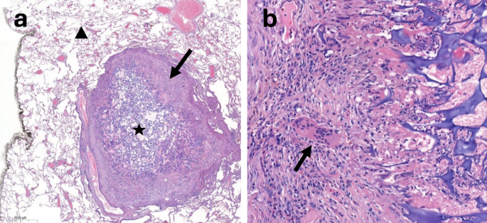
When placed in soft tissues such as lung parenchyma, gelatin sponge is supposed to be absorbed completely within 4 to 6 weeks [13]. However, in this study, 33 PTN remained visible more than 6 weeks after the procedure and in one case the nodule was even still visible on imaging after more than 5 months. Currently, the absorption process, which involves an inflammatory cellular response to foreign material with immune cells such as monocytes, neutrophils, lymphocytes and macrophages, is not fully understood [14]. Absorption time is influenced by various factors, including the site of use, the amount of material used, and the local degree of saturation with blood [15]. Moreover, in case of resistance to degradation by phagocytic cells, foreign body granulomas can occur. It is a specialised form of chronic inflammatory tissue characterised histopathologically by the presence of merging macrophages that form multinucleated giant cells [16, 17]. It has been described with gelatin sponge, in particular in the brain [18, 19]. Formation of a granuloma against gelatin sponge sealant deposited in the lung, as illustrated in Fig. 3, can also explain a prolonged degradation time.
Histopathologic appearance of a pulmonary track nodule 6 weeks after track sealing with gelatin sponge slurry visible on a surgical specimen (hematoxylin and eosin). (a) The acellular gelatin sponge material (star) is surrounded by a ring of non-necrotizing granulomatous tissue containing chronic inflammatory cells (arrow), mainly macrophages, within the pulmonary parenchyma (triangle). (b) Higher magnification of the inflammatory tissue adjacent to the sealant, showing the presence of multinucleated giant cells (arrow)
The issue of the fate of track sealant released in lung parenchyma after CT-guided lung biopsy has already been addressed for hydrogel plugs, a manufactured sealant made of biodegradable synthetic polymer [20, 21]. Recently, Moussa et al. assessed the radiological aspect of hydrogel plugs released to the lung parenchyma on cross-sectional chest imaging performed after CT-guided lung biopsy [22]. They observed the appearance of linear, serpiginous or lobulated opacities, extending from the pleural surface to the lesion, which were for some of them hypermetabolic on [18F]FDG PET/CT. In a similar study, De Groot et al. drew attention to the fact that the persistence of a visible track within the lung parenchyma on imaging after TS with hydrogel plugs can mimic malignant cell dissemination along the biopsy needle and could complicate target delineation before stereotactic body radiation therapy [23].
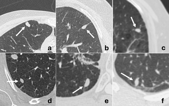
Unlike hydrogel plugs, TS with GSS resulted in nodular opacities, probably due to the more fluid and dispersible characteristics of GSS compared to the elongated shape of the plugs. The nodular appearance may lead to a higher risk of radiological misinterpretation as it could be more easily mistaken with a new tumoral lesion, especially as the interval time between the biopsy and the follow-up CT is long. However, in this study, the shape of PTNs was characteristically ovoid, with smooth borders and well-defined margins, which are radiological features that can help differentiate PTN from tumoral nodule (Fig. 4). Moreover, the risk of malignant cells deposited along the track of a needle during a biopsy resulting in a new nodule is low, especially when a coaxial needle is used [24,25,26]. This risk is particularly low in patients with intrapulmonary malignancy, contrary to pleural neoplasm [27].
Besides the type of sealant, the length of the pulmonary track was another factor associated with a higher risk of PTN in this study: longer tracks were more frequently associated with PTN (p = .009). This could be explained by technical differences, including a likely lower volume of sealant released in shorter pulmonary tracks.
Despite the inflammatory reaction typically associated with the absorption process, 86% of the PTNs were not hypermetabolic on [18F]FDG PET/CT when available. This may be due to a low number of inflammatory cells and/or the small size of the nodules, which might be below the spatial resolution of PET. Nevertheless, [18F]FDG PET/CT might be useful for differentiating PTNs from new tumoral lesions, even if the small size of the nodules may limit its sensitivity.
In this study, two PTNs have also been demonstrated in the group of patients who received no embolization. After a review of the cases, both patients were found to have extensive alveolar haemorrhage along the biopsy track, and PTN in these cases may correspond to residual hematomas. No PTN was detected in the saline group.
This study has several limitations. First, this is a retrospective study. Consequently, the time between the biopsy and the follow-up chest CT varied significantly among patients, and was significantly shorter in the no TS group compared to the GSS and saline groups (p = .002). Therefore, since PTN are absorbed over time, they may have been under detected in the GSS and saline groups. Furthermore, due to the lack of systematic and planned follow-up CTs, the time during which the PTNs were visible was difficult to estimate precisely. We therefore decided to report the last date when the nodule was clearly visible, leading to an underestimation of the true lasting time of these nodules. Finally, the presence of a PTN after TS is undoubtedly related to some technical parameters of the procedure (i.e., the volume of sealant injected, the pressure applied during injection of sealant, the needle withdrawal speed), and may therefore depend on the operators, as shown in our results.
In conclusion, this study has demonstrated that TS with GSS after CT-guided lung biopsy commonly resulted in a visible nodule along the pulmonary track on subsequent imaging. These nodules typically have smooth borders and ovoid shapes, and should not be mistaken with tumor cell dissemination along the biopsy needle track.
Data availability
The datasets used and/or analysed during the current study are available from the corresponding author on reasonable request.
Abbreviations
- TS:
-
Track sealing
- GSS:
-
Gelatin sponge slurry
- PTN:
-
Pulmonary track nodule
- [18F]FDG:
-
[18F]fluoro-2-deoxy-2-D-glucose
- PET/CT:
-
Positron emission tomography/CT
- SUVmax :
-
Maximum standardized uptake value
- OR:
-
Odds ratio
- CI:
-
Confidence intervals
References
Heerink WJ, De Bock GH, De Jonge GJ, Groen HJM, Vliegenthart R, Oudkerk M. Complication rates of CT-guided transthoracic lung biopsy: meta-analysis. Eur Radiol. 2017;27(1):138–48.
Lokhandwala T, Bittoni MA, Dann RA, D’Souza AO, Johnson M, Nagy RJ, et al. Costs of diagnostic assessment for lung cancer: A medicare claims analysis. Clin Lung Cancer. 2017;18(1):e27–34.
Vachani A, Zhou M, Ghosh S, Zhang S, Szapary P, Gaurav D, et al. Complications after transthoracic needle biopsy of pulmonary nodules: A Population-Level retrospective cohort analysis. J Am Coll Radiol. 2022;19(10):1121–9.
Huo YR, Chan MV, Habib AR, Lui I, Ridley L. Post-Biopsy manoeuvres to reduce pneumothorax incidence in CT-Guided transthoracic lung biopsies: A systematic review and Meta-analysis. Cardiovasc Intervent Radiol. 2019;42(8):1062–72.
Grange R, Di Bisceglie M, Habert P, Resseguier N, Sarkissian R, Ferre M, et al. Evaluation of preventive tract embolization with standardized gelatin sponge slurry on chest tube placement rate after CT-guided lung biopsy: a propensity score analysis. Insights Imaging. 2023;14(1):212.
Renier H, Gérard L, Lamborelle P, Cousin F. Efficacy of the tract embolization technique with gelatin sponge slurry to reduce pneumothorax and chest tube placement after percutaneous CT-guided lung biopsy. Cardiovasc Intervent Radiol. 2020;43(4):597–603.
Sum R, Lau T, Paul E, Lau K. Gelfoam slurry tract occlusion after computed tomography-guided percutaneous lung biopsy: does it prevent major pneumothorax? J Med Imaging Radiat Oncol. 2021;65(6):678–85.
Tran AA, Brown SB, Rosenberg J, Hovsepian DM. Tract embolization with gelatin sponge slurry for prevention of pneumothorax after percutaneous computed Tomography-Guided lung biopsy. Cardiovasc Intervent Radiol. 2014;37(6):1546–53.
Baadh AS, Hoffmann JC, Fadl A, Danda D, Bhat VR, Georgiou N, et al. Utilization of the track embolization technique to improve the safety of percutaneous lung biopsy and/or fiducial marker placement. Clin Imaging. 2016;40(5):1023–8.
Dheur S, Gérard L, Lamborelle P, Valkenborgh C, Grandjean F, Gillard R et al. Track sealing in CT-guided lung biopsy using gelatin sponge slurry versus saline in reducing post-biopsy pneumothorax: a prospective randomised study. J Vasc Interv Radiol. 2024;S1051044324004949.
Abada HT, Golzarian J. Gelatine sponge particles: handling characteristics for endovascular use. Tech Vasc Interv Radiol. 2007;10(4):257–60.
Bankier AA, MacMahon H, Colby T, Gevenois PA, Goo JM, Leung ANC, et al. Fleischner society: glossary of terms for thoracic imaging. Radiology. 2024;310(2):e232558.
Jenkins HP. Gelatin sponge, a new hemostatic substance: studies on absorbability. Arch Surg. 1945;51(4):253.
Das D, Zhang Z, Winkler T, Mour M, Günter CI, Morlock MM et al. Bioresorption and Degradation of Biomaterials. In: Kasper C, Witte F, Pörtner R, editors. Tissue Engineering III: Cell - Surface Interactions for Tissue Culture [Internet]. Berlin, Heidelberg: Springer Berlin Heidelberg; 2011 [cited 2024 Apr 18]. pp. 317–33. (Advances in Biochemical Engineering/Biotechnology; vol. 126). Available from: https://doiorg.publicaciones.saludcastillayleon.es/10.1007/10_2011_119
Gelfoam instruction for use [Internet]. Available from: https://cdn.pfizer.com/pfizercom/products/uspi_gelfoam_plus.pdf
Williams DF. Tissue-biomaterial interactions. J Mater Sci. 1987;22(10):3421–45.
Shah KK, Pritt BS, Alexander MP. Histopathologic review of granulomatous inflammation. J Clin Tuberc Mycobact Dis. 2017;7:1–12.
Knowlson GTG. Gel-foam granuloma in the brain. J Neurol Neurosurg Psychiatry. 1974;37(8):971–3.
Guerin C, Heffez DS. Inflammatory intracranial mass lesion: an unusual complication resulting from the use of gelfoam. Neurosurgery. 1990;26(5):856–9.
Butnor KJ, Bodolan AA, Bryant BRE, Schned A. Impact of histopathologic changes induced by polyethylene glycol hydrogel pleural sealants used during transthoracic biopsy on lung cancer resection specimen staging. Am J Surg Pathol. 2020;44(4):490–4.
Yousem SA, Amin RM, Levy R, Baker N, Lee P. Pulmonary pathologic alterations associated with biopsy inserted hydrogel plugs. Hum Pathol. 2019;89:40–3.
Moussa AM, Hui Y, Araujo Filho JA, Muallem N, Li D, Jihad M, et al. Radiologic and histopathologic features of hydrogel sealant after lung resection in participants of a prospective randomized clinical trial. Clin Imaging. 2023;95:92–6.
De Groot PM, Shroff GS, Ahrar J, Sabloff BS, Gladish GM, Moran C, et al. Implications for high-precision dose radiation therapy planning or limited surgical resection after percutaneous computed tomography-guided lung nodule biopsy using a tract sealant. Adv Radiat Oncol. 2018;3(2):139–45.
Ibukuro K, Tanaka R, Takeguchi T, Fukuda H, Abe S, Tobe K. Air embolism and needle track implantation complicating CT-guided percutaneous thoracic biopsy: single-institution experience. AJR Am J Roentgenol. 2009;193(5):W430–436.
Tomiyama N, Yasuhara Y, Nakajima Y, Adachi S, Arai Y, Kusumoto M, et al. CT-guided needle biopsy of lung lesions: A survey of severe complication based on 9783 biopsies in Japan. Eur J Radiol. 2006;59(1):60–4.
Ayar D, Golla B, Lee JY, Nath H. Needle-Track metastasis after transthoracic needle biopsy. J Thorac Imaging. 1998;13(1):2–6.
Robertson EG, Baxter G. Tumour seeding following percutaneous needle biopsy: the real story! Clin Radiol. 2011;66(11):1007–14.
Acknowledgements
Not applicable.
Funding
This research did not receive any specific grant from funding agencies in the public, commercial, or not-for-profit sectors.
Author information
Authors and Affiliations
Contributions
Study conception and design: FG and FC; data generation and collection: NW, ND, LG, PL, LG, CV; statistical analyses: ND; interpretation of results: FC, FG, ND; draft manuscript preparation: FC, FG. All authors reviewed the results and approved the final version of the manuscript.
Corresponding author
Ethics declarations
Ethics approval and consent to participate
The study was conducted in accordance with the Declaration of Helsinki. The institutional review board (Comité d’Ethique Hospitalo-Facultaire Universitaire de Liège, CHU de Liège) approved this retrospective study, and the need for informed consent was waived based on its retrospective design (date: 1st of October 2024; Reference number: 2024/351).
Consent for publication
Not applicable.
Competing interests
The authors declare no competing interests.
Additional information
Publisher’s note
Springer Nature remains neutral with regard to jurisdictional claims in published maps and institutional affiliations.
Rights and permissions
Open Access This article is licensed under a Creative Commons Attribution-NonCommercial-NoDerivatives 4.0 International License, which permits any non-commercial use, sharing, distribution and reproduction in any medium or format, as long as you give appropriate credit to the original author(s) and the source, provide a link to the Creative Commons licence, and indicate if you modified the licensed material. You do not have permission under this licence to share adapted material derived from this article or parts of it. The images or other third party material in this article are included in the article’s Creative Commons licence, unless indicated otherwise in a credit line to the material. If material is not included in the article’s Creative Commons licence and your intended use is not permitted by statutory regulation or exceeds the permitted use, you will need to obtain permission directly from the copyright holder. To view a copy of this licence, visit http://creativecommons.org/licenses/by-nc-nd/4.0/.
About this article
Cite this article
Grandjean, F., Withofs, N., Detrembleur, N. et al. Incidence and features of pulmonary track nodules after CT-guided lung biopsy with track sealing using gelatin sponge slurry. BMC Med Imaging 25, 107 (2025). https://doiorg.publicaciones.saludcastillayleon.es/10.1186/s12880-025-01644-x
Received:
Accepted:
Published:
DOI: https://doiorg.publicaciones.saludcastillayleon.es/10.1186/s12880-025-01644-x


