- Research
- Open access
- Published:
The impact of cerebral vessels morphological alteration and white matter hyperintensities burden on the one-year risk of ischemic stroke recurrence
BMC Medical Imaging volume 25, Article number: 150 (2025)
Abstract
Purpose
To assess the associations between cerebral vessels morphological features, white matter hyperintensities (WMHs), and the one-year risk of ischemic stroke recurrence.
Methods
A total of 677 patients diagnosed with acute ischemic stroke from January 2018 to April 2021 were consecutively enrolled. Head computed tomography angiography (CTA) and magnetic resonance imaging including fluid-attenuated inversion recovery (FLAIR), were obtained on admission. Cerebral vessels morphological features such as volume, length, radius, density, tortuosity, branch complexity, and degree of stenosis were extracted and calculated using CTA data. Additionally, automated segmentation was employed for delineating WMHs lesions based on FLAIR images. By incorporating clinical characteristics, six predictive models were developed using Cox proportional hazards analysis to estimate the one-year risk of stroke recurrence. The performance of these models was evaluated by comparing the concordance index (C-index).
Results
The study found significant associations between the lack of antiplatelet therapy at discharge, reduced length and branching of cerebral vessels, and increased burden of WMHs, with a higher one-year risk of recurrent ischemic stroke (all P < 0.05). The integrated model demonstrated superior prognostic capability (C-index: 0.750; 95% CI: 0.684–0.817), outperforming models based solely on clinical characteristics (C-index: 0.636; 95% CI: 0.555–0.717), cerebral vessels morphology (C-index: 0.601; 95% CI: 0.526–0.676), and WMHs burden (C-index: 0.680; 95% CI: 0.603–0.757).
Conclusion
The quantitative assessment of cerebral vessels morphological features and WMHs provides a promising neuroimaging tool for estimating the one-year risk of ischemic stroke recurrence. The incorporation of cerebral vessels morphological features enhances the predictive accuracy.
Introduction
Stroke is a prominent contributor to mortality and morbidity on a global scale, serving as a significant catalyst for cognitive impairment and functional disability [1, 2]. It is estimated that ischemic strokes account for approximately 87% of all stroke cases [3]. Though significant progress has been made in the individualized antithrombotic therapy, standardized risk factors controlling and stenting in the secondary prevention for ischemic stroke, there remains a persistent risk of stroke recurrence, with 1.9–6% within the first 3 months and 7.2–8.0% within the first one year. A randomized, double-blind, placebo-controlled trial [4] was conducted at 202 centers in China involving patients with a minor ischemic stroke or transient ischemic attack (TIA) who carried CYP2C19 loss-of-function alleles. Stroke occurred within 90 days in 191 patients (6.0%) in the ticagrelor group and 243 patients (7.6%) in the clopidogrel group. An investigator-initiated, open-label trial [5] at 103 sites in 15 countries demonstrated that recurrent ischemic stroke occurred in 14 participants (1.4%) in the early initiation of direct oral anticoagulants group and 25 participants (2.5%) in the later initiation of direct oral anticoagulants group by 30 days and in 18 participants (1.9%) and 30 participants (3.1%), respectively, by 90 days. Recurrent strokes are widely acknowledged as being more frequently associated with fatality and disability compared to initial events [6, 7]. Therefore, recognizing other factors influencing stroke recurrence is warranted to provide new management strategy for reducing the relapse of ischemic stroke.
A recent investigation demonstrated that decreased arterial branch density, increased mean radius and mean tortuosity were associated with aging in population aged 45 to 54 years after adjusting for vascular risk factors, suggesting the potential significance of early preservation of cerebrovascular morphologies for preventing cerebrovascular disease [8]. Zhang et al. [9] demonstrated that arterial tortuosity is associated with spontaneous cervicocerebral artery dissection, a significant cause of ischemic stroke that frequently occurs in young and middle-aged patients. Similarly, previous studies have shown the relationships between geometric features of intracranial arteries and the distribution and burden of atherosclerotic plaques, revealing the importance of cerebrovascular morphologies in the development of ischemic stroke [10, 11]. Hence, it is convinced to hypothesize that there are associations between cerebrovascular morphologies and the recurrence of ischemic stroke, where the morphological features of cerebral vessels include vascular volume fraction, branch point density, vessels diameter, length, and tortuosity. Besides, a meta-analysis including 104 studies has shown that moderate-severe white matter hyperintensities (WMHs) at baseline increased the risk of recurrent ischemic stroke [12], and increased WMHs lesion volume was reported to be associated with reduced vessels density and tortuosity, as well as enlarged radius [13]. There is evidence linking cerebrovascular morphological changes to WMHs, as demonstrated in studies involving young adults where WMHs lesion counts were found to be associated with cerebral vessels densities [13, 14]. Nevertheless, the specific link between cerebral vessels morphology alterations and WMHs in stroke patients, as well as the comprehensive relationship among WMHs, cerebrovascular vessels morphology alterations, and stroke recurrence, has not been researched.
Hence, this investigation aims to explore the relationships between cerebrovascular morphological alterations, WMHs, and the recurrence of strokes. By conducting a one-year follow-up study on patients who have suffered an ischemic stroke, a predictive model that combines both cerebrovascular characteristics and WMHs was aimed to develop to assess the risk of one-year stroke recurrence.
Materials and methods
Study population
This study was granted approval by the Institutional Ethics Committee of Minhang Hospital, Fudan University, and the requirement for obtaining informed consent was waived (approval number: 2022-013-01 K). The study adhered to the principles outlined in the 1964 Declaration Helsinki and its subsequent revisions.
This study included patients with first-ever ischemic stroke at our stroke center between January 2018 and April 2021. Inclusion criteria were as follows: age ≥ 18, ischemic stroke diagnosed by both clinical symptoms and imaging findings, with an onset within two weeks. Exclusion criteria encompassed non-first ever stroke patients, pre-existing neurological or psychiatric disorders, cerebral tumors, history of substance abuse, severe MRI (magnetic resonance imaging, MRI) artifacts, CTA artifacts, mortality during follow-up or loss to follow-up.
Clinical data
Demographic and clinical characteristics were collected, including age, sex, smoking status, alcohol consumption, presence of hypertension, hyperlipidemia, diabetes mellitus and atrial fibrillation. Clinical assessments included the National Institutes of Health Stroke Scale (NIHSS) scores at admission, the Trial of Org 10,172 in Acute Stroke Treatment (TOAST) classification [15], acute interventions (thrombolysis, embolectomy), and medications prescribed post-discharge (antiplatelets, anticoagulants, statins). Outcomes were measured using the modified Rankin Scale (mRS) at 90 days post-stroke. Smoking status was defined as current (≥ 1 cigarette/day in past year) or former smoker. Alcohol consumption was defined as current (≥ 12 g ethanol/day) or former drinker. Hypertension: systolic BP ≥ 140 mmHg and/or diastolic BP ≥ 90 mmHg, or antihypertensive medication use. Diabetes mellitus: fasting glucose ≥ 7.0 mmol/L, HbA1c ≥ 6.5%, or hypoglycemic therapy. Hyperlipidemia: total cholesterol ≥ 6.2 mmol/L, LDL ≥ 4.1 mmol/L, or lipid-lowering medication. A study flow chart detailing these components is depicted in Fig. 1.
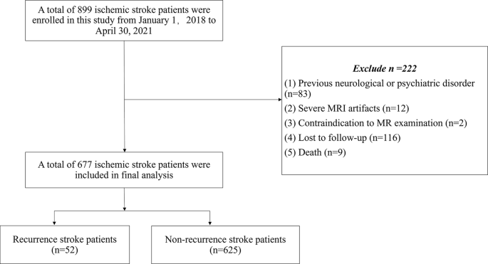
Study population flowchart
Head CTA acquisition
A comprehensive CTA protocol was employed using the SOMATOM Force CT scanner (Siemens Medical Solutions), involving NCCT and CTA of the head and neck. A contrast agent (320 mg iodine/mL; Jiangsu Hengrui Medicine Co., Ltd., Jiangsu, China) was administered intravenously, followed by a saline flush, with parameters adjusted for optimal imaging quality as detailed in the protocol.
Brain MRI acquisition
MRI data were acquired using two scanners: the EXCITE HD 1.5 T MRI (GE Healthcare) and the uMR780 3.0 T MRI (United Imaging Healthcare). The protocol comprised axial T2-weighted imaging, T1-weighted imaging (T1-WI), T2FLAIR (fluid-attenuated inversion recovery), and diffusion-weighted imaging (DWI). Detailed scan parameters are provided in Table 1.
Outcome assessment
Recurrent ischemic stroke was defined as the emergence of a novel neurological deficit or deterioration of an existing neurological deficit accompanied by newly identified discrete lesions on DWI [16]. All instances of ischemic stroke recurrence were confirmed at our stroke center or documented in diagnostic reports from other stroke centers. We assessed the mRS at 90 days and documented stroke recurrence within one year after the qualifying event in-person or by telephone interviews with patients or their caregivers.
Cerebral vessels segmentation
Cerebrovascular segmentation was performed on head CTA image following these steps: (1) Enhancement of blood vessels structures was achieved using the Jerman enhancement filter [13, 17], which advanced the multi-scale Hessian filtering with a novel enhancement function and promoted better and uniformed responses for vessels of different sizes and varying contrast, e.g. at bifurcations; (2) The enhanced vascular image was then binarized as the initial segmentation mask for CTA, level-set method, which allowed multi-object segmentation in one plane, was conducted to obtain the accurate boundary of vessels for each CTA slice; (3) The segmentation outcomes were preliminarily evaluated by two experienced neuroradiologists to configure batch processing parameters, followed by a thorough visual inspection to identify and correct deficiencies such as insufficient vascular branches or non-vascular tissue inclusions. (4) Final adjustments were made manually using ITK-SNAP software (http://www.itk-snap.org), ensuring accuracy in the segmented cerebral vessels images (All cerebral vessels segmentation details were shown in Fig. 2).

Cerebral vessels segmentation pipeline. The Jerman enhancement filter highlighted blood vessels structures. An automatic segmentation technique then merged the CTA data with the enhanced images using a level-set-based approach. ITK-SNAP software verified the accuracy of the segmented images. The segmentation results of intracranial blood vessels were integrated with brain masks to calculate vascular volume and cerebral vascular density. Finally, the Skeleton 3D toolbox extracted the centerlines of blood vessels, enabling accurate vessels length calculation
Cerebral vessels morphology features extraction
The segmentation results of intracranial blood vessels were obtained by combining the segmentation results of blood vessels with brain masks, enabling direct calculation of vascular volume and cerebral vascular density. The Skeleton 3D toolbox (Skeleton3D Calculates the 3D skeleton of an arbitrary binary volume using parallel medial axis thinning. Detailed code is available at: https://github.com/phi-max/skeleton3d-matlab) was utilized to extract the centerline of blood vessels from the segmentation results, facilitating the calculation of vessels length feature. Morphological features including cerebral vessels volume, length, radius, density, tortuosity, and branching were quantified. Cerebral vascular density is defined as the ratio between segmented intracranial arterial volume from CTA and brain volume. Cerebral vessels radius represents the distance from a point on the centerline to the outermost boundary of blood vessels, where average radius denotes an overall calculation for all points on the centerline. Cerebral vessels tortuosity quantifies twisting through a ratio between shortest path length and actual length. Blood vessels branch number refers to bifurcation points in blood vessels (Definition of Cerebral Vessels Morphology Features was shown in Fig. 3). Each individual’s radius distribution (or histogram) comprises radius values at all points along their respective centerlines. All data processing was performed on MATLAB (version 2019a, MathWorks, Natick, Massachusetts, USA).

Definition of Cerebral Vessels Morphology Features. (1) Cerebral Vessels Density: Ratio of segmented intracranial arterial volume (derived from CTA) to total brain volume. (2) Vessel Radius: Distance from the vessel centerline to the outermost boundary. Average radius is computed as the mean value across all centerline points. (3) Vessel Tortuosity: Ratio of the shortest path length to the actual vessel length. (4) Branch Number: Total count of bifurcation points in the vascular network. Note: CTA = Computed Tomography Angiography. Schematic diagram created by the authors to illustrate morphological feature definitions and do not represent specific patient data
WMHs segmentation and extraction
We employed an automated deep learning-based pipeline for WMH segmentation and extraction, implemented as follows:
Our study utilizes the nnU-Net deep learning framework (https://github.com/MIC-DKFZ/nnUNet) [18], nnU-Net is a state-of-the-art, self-configuring framework for biomedical image segmentation that automates the entire pipeline, including preprocessing, network architecture design, training, and postprocessing. Then, we trained with manually annotated T1-WI and FLAIR images from our previous studies [19, 20]. The trained model is subsequently employed for the segmentation and extraction of white matter hyperintensities in our study. Furthermore, by referencing the brain region divisions in the UBO Detector [21], we obtain the periventricular (PV-WMHs) and deep white matter hyperintensities (D-WMHs), as well as the white matter hyperintensities in the frontal, temporal, parietal, occipital lobes, brainstem and cerebellum. A distance threshold of 10 mm from the ventricular border was employed to identify PV-WMHs and deep white matter hyperintensities D-WMHs [21]. The volumes of PV-WMHs (PV-WMHV), D-WMHs(D-WMHV), and WMHs in different lobes were quantified. In cases where one hemisphere was affected by infarct lesions that also exhibited hyperintensities, we referred to the contralateral hemisphere to minimize confusion. A total of 14 WMHV parameters were obtained (Table 2).
The study acquired a comprehensive set of six vessels morphology features, encompassing volume, length, radius, density, tortuosity, and branching. Moreover, the degree of stenosis in the offending vessels on the same hemisphere as the infarction lesion (including segments C1-C7 of the internal carotid artery, A1-A3 of the anterior cerebral artery, M1-M2 of the middle cerebral artery, and P1-P2 of the posterior cerebral artery) was evaluated by two neuroradiologists. The stenotic vessels were categorized into 4 groups by the grade of stenosis: normal (<30%), mild (30–49%), moderate (50–69%), or severe (≥ 70%) [22].
Statistical analysis
The statistical analysis was performed with R software (version 3.6.2, http://www.R-project.org). The rms package was used for Cox proportional hazards regression, nomograms, and decision curve analysis (DCA). The Hmisc package was used for comparisons between C-indexes. The reported statistical significance levels were all two-sided, with a P-value < 0.05 representing statistical significance.
We applied Spearman correlation for evaluating relationships between WMHs volume and cerebral vessels features. Group differences based on recurrent stroke presence were assessed via the Mann-Whitney U and chi-square or Fisher’s exact tests as appropriate. All continuous variables, including WMHV, vessels morphology features and age, were classified into high-risk or low-risk groups according to stroke recurrence event by using Kaplan-Meier survival analysis, whose optimal threshold was identified corresponding to the minimum P-value. Multivariate Cox hazards regression (backward step-down selection) was used to select the features and construct the prediction model. The clinical characteristics model (model 1), the cerebral vessels morphology features model (model 2), the clinical characteristics combined with vessels morphology features model (model 3), the quantitative WMHs model (model 4), the clinical characteristics combined with WMHs model (model 5), and the integrated model of WMHs, cerebral vessels and clinical characteristics (model 6) were ultimately constructed.
The performance of the six different models was compared. The Harrell concordance index (C-index) was calculated to demonstrate the discrimination performance, along with the concordance probability estimate considering the high degree of censoring in our data (1 indicates perfect concordance; 0.5 indicates no better concordance than chance) [23]. The optimal prediction model nomogram was presented. Additionally, a DCA was employed to assess the clinical utility of the predictive models.
Results
Demographic and clinical characteristics
Among the 899 subjects with ischemic stroke, 222 were excluded from the study because of the following reasons: (1) previous neurological or psychiatric disorders (n = 83); (2) severe MRI artifacts (n = 12); (3) contraindication to MR examination (n = 2); (4) lost to follow-up (n = 116) and (5) death (n = 9).
Consequently, a total of 677 patients were included in the final analysis, with a mean age of 65.4 ± 11.46 years. Among them, 52 patients (7.7%) experienced recurrence of ischemic stroke within one year after the qualifying event. Regarding the subtype of ischemic stroke, the one-year recurrence rates for large artery atherosclerosis (LAA), cardioembolic (CE), small vessels occlusion (SVO), other causes, and undetermined causes were 9.4%, 12.2%, 5.2%, 0%, and 6.8% respectively. Patients with recurrent stroke (mean age: 67.8 ± 10.96) were older than those without recurrence (mean age: 65.22 ± 11.486). There were no significant differences observed between the recurrence and non-recurrence groups in terms of smoking status, alcohol consumption, presence of hypertension, hyperlipidemia, diabetes mellitus, atrial fibrillation, NIHSS score on admission, stroke subtype, mRS at 90 days or anticoagulants administered post-discharge (Table 2). However, we did find that lack of antiplatelets administered post-discharge was associated with a higher risk of recurrence (P < 0.05) (Table 2).
The correlation between WMHV and morphological features of cerebral vessels
The analysis revealed a negative correlation between WMHV in various spatial distributions and the volume, length, density, and branching of cerebral vessels. Conversely, cerebral vessels tortuosity and radius showed a positive correlation with WMHV. (Fig. 4).

Heatmap of the correlation between cerebral vessels morphologic features and white matter hyperintensities volume varying spatial distribution. The color code indicated the correlation. The embedded text showed the P value. The asterisk (*) indicated P value < 0.05. W1: the whole brain white matter hyperintensities volume (WMHV); W2: periventricular WMHV; W3: deep WMHV; W4: left frontal lobe WMHV; W5; right frontal lobe WMHV; W6: left temporal lobe WMHV; W7: right temporal lobe WMHV; W8: left parietal lobe WMHV; W9: right parietal lobe WMHV; W10: left occipital lobe WMHV; W11: right occipital lobe WMHV; W12: left cerebellum WMHV; W13: right cerebellum WMHV; W14: brainstem WMHV
Prediction model construction
Six prediction models were constructed to assess stroke recurrence risk, with their performances detailed in Table 3. Survival curves for the models are depicted in Fig. 5.

Kaplan-Meier survival curves analysis according to six different prediction models for patients with ischemic stroke. A significant association of the six prediction models with the disease-free survival was shown in the graph (All P < 0.05 by log-rank test). (A) The clinical characteristics model (model 1), (B) the cerebral vessels morphology features model (model 2), (C) the clinical characteristics combined with vessels morphology features model (model 3), quantitative WMHs-based model in training cohort, (D) the quantitative WMHs model (model 4). (E) the clinical characteristics combined with WMHs model (model 5), (F) the integrated model of WMHs, cerebral vessels and clinical characteristics (model 6)
Model 1 utilized multiple Cox proportional hazard regression analysis, revealing that post-discharge antiplatelet use was significantly associated with a reduced stroke recurrence risk (Hazard Ratio [HR] = 0.323, 95% Confidence Interval [CI]: 0.151–0.691, P = 0.004).
Model 2 indicated, through similar analysis, that an increased number of cerebral vessels branches was significantly associated with a lower risk of recurrence (HR = 0.921, 95% CI: 0.861–0.986, P = 0.018).
In Model 3, antiplatelet therapy post-discharge (HR = 0.333, 95% CI: 0.154–0.721, P = 0.005) and a higher number of cerebral vessels branches (HR = 0.910, 95% CI: 0.848–0.976, P = 0.008) were identified as significant predictors for reduced stroke recurrence risk.
Model 4 findings, through multiple Cox regression, demonstrated significant associations of D-WMHV (HR = 0.696, 95% CI: 0.548–0.885, P = 0.003) and WMHV in various brain regions with recurrence risk, highlighting specific areas like the right frontal (HR = 1.046, 95% CI: 1.000-1.094, P = 0.048) and bilateral parietal lobes (right parietal lobes: HR = 1.128, 95% CI: 1.049–1.213, P = 0.031; left parietal lobes: HR = 1.058, 95% CI: 1.005–1.113, P = 0.001) as significant factors.
In Model 5, stroke subtype (HR = 0.759, 95% CI: 0.593–0.972, P = 0.029), post-discharge antiplatelet therapy (HR = 0.421, 95% CI: 0.204–0.870, P = 0.019), and specific WMHV metrics like D-WMHV (HR = 0.646, 95% CI: 0.501–0.834, P = 0.001) and WMHV in frontal and parietal lobes(right frontal lobes: HR = 1.055, 95% CI: 1.008–1.105, P = 0.021; right parietal lobes: HR = 1.077, 95% CI: 1.020–1.136; left parietal lobes: HR = 1.162, 95%CI: 1.073–1.260, all P < 0.05) were found to significantly influence recurrence risk.
Model 6 integrated several variables including cerebral vessels length (HR = 0.943, 95%CI:0.895–0.992, P = 0.024), stroke subtype (HR = 0.757, 95% CI: 0.592–0.967, P = 0.026), antiplatelet therapy post-discharge (HR = 0.311, 95% CI: 0.142–0.680, P = 0.003), and WMHV in multiple brain regions (D-WMHV[HR = 0.660, 95% CI: 0.510–0.853, P = 0.002], right frontal WMHV[HR = 1.054, 95% CI: 1.006–1.104, P = 0.027], bilateral parietal lobe WMHV [right parietal: HR = 1.074, 95% CI: 1.018–1.133; left parietal: HR = 1.147, 95%CI: 1.060–1.243, all P < 0.05]). This model illustrated significant associations with recurrence risk.
Incremental value of cerebral vessels morphology features and WMHs in stroke recurrence prediction
The nomogram was generated combining clinical characteristics, cerebral vessels morphology features, WMHV (Fig. 6). C-indexes for the different models are listed in Table 3. The integrated model, demonstrated superior prognostic capability, achieving a higher C-index than models based solely on clinical characteristics, cerebral vessels morphology, or WMHs. The DCA illustrates the integrated nomogram has a higher overall net benefit in predicting one-year risk of ischemic stroke recurrence compared other predictive models.

Nomogram developed with decision curve analysis. (A) A nomogram was developed with clinical characteristics, cerebral vessels morphological features and white matter hyperintensities volume. (B) The net benefit was calculated by summing the benefits (true positive results) and subtracting the harms (false-positive results). The model (purple line) showed higher net benefit compared with other models
Discussion
The results of this study underscore the significant correlation between clinical characteristics, cerebral vessels morphology, and WMHV, and their combined impact on the risk of stroke recurrence. The findings suggest that the model based on quantitative WMHs data significantly outperforms those based solely on clinical or cerebral vessels morphological factors in predicting one-year stroke recurrence risk. The inclusion of cerebral vessels morphological features increases the model’s predictive accuracy for one-year stroke recurrence risk assessment.
A systematic review and meta-analysis [24], encompassing 26 studies conducted between 1997 and 2019, revealed that the recurrence rate of stroke varied from 5.7 to 51.3%. Recurrent stroke was predominantly observed in cases of LAA and CE strokes. The one-year recurrent rate for all acute ischemic stroke subtypes within this population was found to be consistent with previous research at 7.7%. Furthermore, our study also identified LAA and CE as the most prevalent stroke subtypes. This study also demonstrates that adherence to antiplatelet therapy post-discharge significantly reduces the risk of stroke recurrence. This is in line with previous research indicating an inverse relationship between antiplatelet therapy at discharge and stroke recurrence rates [25]. It is hypothesized that non-adherence to antiplatelet therapy for secondary prevention is associated with an increased risk of ischemic stroke recurrence.
WMHs of presumed vascular origin are the most common neuroimaging feature of small vessels disease [26]. WMHs may manifest as superficial manifestations of cerebrovascular disease, while their underlying mechanism involves pathological changes in cerebral vessels, leading to morphological alterations. The observed variance in WMHs, reflecting distinct underlying cerebral parenchymal changes, may be linked to cerebrovascular morphological alterations, as supported by evidence from a study in young adults linking WMHs counts to cerebral vessels density [13, 14]. Our findings confirm the association between WMHs and various morphological characteristics of cerebral vessels, indicating that changes in vessels volume, length, density, branching, and tortuosity correlate with WMHs burden. Previous research in young adults without clinical signs of cerebrovascular disease showed an association between an increased number of modifiable cardiovascular risk factors and higher cerebral vessels density and caliber [14]. A study demonstrated the increase in age was found negatively associated with number of branches while positively associated with tortuosity, which remained after adjusting for cardiovascular risk factors [27]. Li et al. [28], revealed that mean length of lenticulostriate artery (LSA), mean tortuosity of LSAs, dilated LSAs, and normalized wall index are associated with large single subcortical infarction. The inverse relationship between branch vessels density and recurrent stroke risk may reflect the role of pial collateral networks. Enhanced branching patterns and vessels density on CTA could indicate robust collateral circulation, which has been shown to preserve perfusion during ischemic events [29]. These collaterals potentially mitigate stroke recurrence by providing alternative blood flow pathways, thereby reducing ischemic burden. While our study did not directly measure collaterals, prior evidence supports this mechanistic link [30]. Further research using dynamic angiographic techniques is warranted to confirm this hypothesis. A recent study utilizing a mouse model of bilateral common carotid artery stenosis (BCAS) noted a significant reduction in vessels length density and volume fraction within the damaged white matter region of BCAS mice [31]. Additionally, mouse models of chronic hypoperfusion have demonstrated that cerebrovascular alterations can lead to white matter damage and cognitive deficits, supporting the hypothesis that cerebral vessels morphology may influence cerebrovascular event outcomes through its association with WMHV.
A recent large-scale study involving over 7000 patients convincingly demonstrated that the burden of WMHs serves as an independent risk factor for stroke recurrence [16], corroborating our findings. The relationship between WMHs and stroke outcomes remains elusive, with current hypotheses suggesting that severe WMHs may contribute to larger infarct volumes and increased susceptibility to acute ischemia [32], disrupt key neuronal networks [33], and reflect systemic vascular risk factors influencing stroke outcomes [34]. Our research further examines the association between stroke recurrence risk and WMHV in different brain regions, affirming the strong links between total WMHV, and specific regional volumes in the right frontal and bilateral parietal lobes, and stroke recurrence risk.
Our study possesses several notable strengths. Cerebral morphological alterations are evidently associated with cerebral events, and the quantitative assessment of cerebral vessels morphology can enhance interpretability. WMHs segmentation was performed using the nnU-Net deep learning framework. Unlike traditional U-Net variants requiring manual hyperparameter tuning, nnU-Net dynamically adapts to new datasets through heuristic rules and empirical optimizations, ensuring robust performance without task-specific modifications. Additionally, we identified WMHs with diverse spatial distributions encompassing periventricular and deep regions as well as various lobes. This comprehensive evaluation combined with complex clinical characteristics provides novel insights into the relationship between these factors and stroke recurrence risk.
The study has several limitations. Firstly, the research was conducted in a single institution, which may limit the generalizability of the findings. External validation with datasets from various stroke centers is necessary to confirm the robustness of our predictive model. Secondly, the exclusion of patients who died, were lost to follow-up, or presented with suboptimal image quality may introduce selection bias. Future studies will aim to mitigate this by conducting more extensive longitudinal follow-ups. Thirdly, the utilized image thickness in FLAIR and T1-WI was 5 mm, potentially compromising segmentation accuracy. While normalization of T1-WI and co-registration with FLAIR sequences were performed to address this, the precision of segmentation would benefit from the employment of 3D T1-weighted imaging and thinner FLAIR sequences for verification. Lastly, while CT angiography is a valuable non-invasive tool for assessing cerebral arteries, it has lower spatial resolution compared to digital subtraction angiography (DSA), particularly for evaluating distal arterial segments and small perforator vessels. This limitation may affect the comprehensive assessment of vascular morphology and collateral circulation, potentially influencing the study’s findings. Future studies incorporating DSA or high-resolution MRI could further validate these results.
In conclusion, the study posits that the lack of antiplatelet therapy post-discharge, decreased cerebral vessels length and branching, along with increased overall and periventricular WMHs burden significantly correlates with an elevated risk of recurrent ischemic stroke within one year. The quantitative analysis of cerebral vascular features and WMHs may provide a new neuroimaging approach for predicting stroke recurrence.
Data availability
The datasets analyzed in this study are available from the corresponding author on request.
Abbreviations
- WMHs:
-
White matter hyperintensities
- CTA:
-
Computed tomography angiography
- FLAIR:
-
Fluid-attenuated inversion recovery
- TIA:
-
Transient ischemic attack
- C-index:
-
Concordance index
- MRI:
-
Magnetic resonance imaging
- NIHSS:
-
National Institutes of Health Stroke Scale
- TOAST:
-
Trial of Org 10172 in Acute Stroke Treatment
- mRS:
-
Modified Rankin Scale
- T1-WI:
-
T1-weighted imaging
- DWI:
-
Diffusion-weighted imaging
- WMHV:
-
WMHs lesion volumes
- PV-WMHs:
-
Periventricular white matter hyperintensities
- D-WMHs:
-
Deep white matter hyperintensities
- PV-WMHV:
-
The volumes of PV-WMHs
- D-WMHV:
-
The volumes of deep white matter hyperintensities
- DCA:
-
Decision curve analysis
- LAA:
-
Large artery atherosclerosis
- CE:
-
Cardioembolic
- SVO:
-
Small vessels occlusion
- HR:
-
Hazard Ratio
- LSA:
-
Lenticulostriate artery
- BCAS:
-
Bilateral common carotid artery stenosis
References
Campbell BCV, Khatri P. Stroke Lancet. 2020;396(10244):129–42.
Sagnier S, Catheline G, Dilharreguy B, Linck PA, Coupe P, Munsch F, Bigourdan A, Debruxelles S, Poli M, Olindo S, et al. Normal-Appearing white matter integrity is a predictor of outcome after ischemic stroke. Stroke. 2020;51(2):449–56.
Saini V, Guada L, Yavagal DR. Global epidemiology of stroke and access to acute ischemic stroke interventions. Neurology. 2021;97(20 Suppl 2):S6–16.
Wang Y, Meng X, Wang A, Xie X, Pan Y, Johnston SC, Li H, Bath PM, Dong Q, Xu A, et al. Ticagrelor versus clopidogrel in CYP2C19 Loss-of-Function carriers with stroke or TIA. N Engl J Med. 2021;385(27):2520–30.
Fischer U, Koga M, Strbian D, Branca M, Abend S, Trelle S, Paciaroni M, Thomalla G, Michel P, Nedeltchev K, et al. Early versus later anticoagulation for stroke with atrial fibrillation. N Engl J Med. 2023;388(26):2411–21.
Skajaa N, Adelborg K, Horvath-Puho E, Rothman KJ, Henderson VW, Thygesen LC, Sorensen HT. Risks of Stroke Recurrence and Mortality After First and Recurrent Strokes in Denmark: A Nationwide Registry Study. Neurology. 2021.
Lackland DT, Roccella EJ, Deutsch AF, Fornage M, George MG, Howard G, Kissela BM, Kittner SJ, Lichtman JH, Lisabeth LD, et al. Factors influencing the decline in stroke mortality: a statement from the American heart association/american stroke association. Stroke. 2014;45(1):315–53.
Zhang B, Yang Z, Li J, Wang B, Shi H, Wang H, Li Y. Modification of cerebrovascular morphologies during different stages of life. J Cereb Blood Flow Metab. 2022;42(11):2151–60.
Zhang L, Liu X, Gong B, Li Q, Luo T, Lv F, Zheng Y, Zheng W, Guo H. Increased internal carotid artery tortuosity is a risk factor for spontaneous cervicocerebral artery dissection. Eur J Vasc Endovasc Surg. 2021;61(4):542–9.
Yu YN, Li ML, Xu YY, Meng Y, Trieu H, Villablanca JP, Gao S, Feng F, Liebeskind DS, Xu WH. Middle cerebral artery geometric features are associated with plaque distribution and stroke. Neurology. 2018;91(19):e1760–9.
Kim BJ, Kim HY, Jho W, Kim YS, Koh SH, Heo SH, Chang DI, Lee YJ. Asymptomatic Basilar artery plaque distribution and vascular geometry. J Atheroscler Thromb. 2019;26(11):1007–14.
Georgakis MK, Duering M, Wardlaw JM, Dichgans M. WMH and long-term outcomes in ischemic stroke: A systematic review and meta-analysis. Neurology. 2019;92(12):e1298–308.
Zhang B, Wang Y, Wang B, Chu YH, Jiang Y, Cui M, Wang H, Chen X. MRI-Based investigation of association between cerebrovascular structural alteration and white matter hyperintensity induced by high blood pressure. J Magn Reson Imaging. 2021;54(5):1516–26.
Williamson W, Lewandowski AJ, Forkert ND, Griffanti L, Okell TW, Betts J, Boardman H, Siepmann T, McKean D, Huckstep O, et al. Association of cardiovascular risk factors with MRI indices of cerebrovascular structure and function and white matter hyperintensities in young adults. JAMA. 2018;320(7):665–73.
Flach C, Muruet W, Wolfe CDA, Bhalla A, Douiri A. Risk and secondary prevention of stroke recurrence: A Population-Base cohort study. Stroke. 2020;51(8):2435–44.
Ryu WS, Schellingerhout D, Hong KS, Jeong SW, Jang MU, Park MS, Choi KH, Kim JT, Kim BJ, Lee J, et al. White matter hyperintensity load on stroke recurrence and mortality at 1 year after ischemic stroke. Neurology. 2019;93(6):e578–89.
Jerman T, Pernus F, Likar B, Spiclin Z. Enhancement of vascular structures in 3D and 2D angiographic images. IEEE Trans Med Imaging. 2016;35(9):2107–18.
Isensee F, Jaeger PF, Kohl SAA, Petersen J, Maier-Hein KH. nnU-Net: a self-configuring method for deep learning-based biomedical image segmentation. Nat Methods. 2021;18(2):203–11.
Zheng G, Fei B, Ge A, Liu Y, Liu Y, Yang Z, Chen Z, Wang X, Wang H, Ding J. U-fiber analysis: a toolbox for automated quantification of U-fibers and white matter hyperintensities. Quant Imaging Med Surg. 2024;14(1):662–83.
Fei B, Cheng Y, Liu Y, Zhang G, Ge A, Luo J, Wu S, Wang H, Ding J, Wang X. Intelligent cholinergic white matter pathways algorithm based on U-net reflects cognitive impairment in patients with silent cerebrovascular disease. Stroke Vasc Neurol; 2024.
Jiang J, Liu T, Zhu W, Koncz R, Liu H, Lee T, Sachdev PS, Wen W. UBO Detector - A cluster-based, fully automated pipeline for extracting white matter hyperintensities. NeuroImage. 2018;174:539–49.
Jeon JS, Sheen SH, Hwang GJ, Kim HC, Kwon BJ. Feasibility of intravenous flat panel detector CT angiography for intracranial arterial stenosis. AJNR Am J Neuroradiol. 2013;34(1):129–34.
Huang Y, Liu Z, He L, Chen X, Pan D, Ma Z, Liang C, Tian J, Liang C. Radiomics signature: A potential biomarker for the prediction of Disease-Free survival in Early-Stage (I or II) Non-Small cell lung Cancer. Radiology. 2016;281(3):947–57.
Kolmos M, Christoffersen L, Kruuse C. Recurrent ischemic Stroke - A systematic review and Meta-Analysis. J Stroke Cerebrovasc Dis. 2021;30(8):105935.
Jing J, Suo Y, Wang A, Zuo Y, Jiang Y, Liu L, Zhao X, Wang Y, Li Z, Li H, et al. Imaging parameters predict recurrence after transient ischemic attack or minor stroke stratified by ABCD(2) score. Stroke. 2021;52(6):2007–15.
Wardlaw JM, Smith EE, Biessels GJ, Cordonnier C, Fazekas F, Frayne R, Lindley RI, O’Brien JT, Barkhof F, Benavente OR, et al. Neuroimaging standards for research into small vessel disease and its contribution to ageing and neurodegeneration. Lancet Neurol. 2013;12(8):822–38.
Chen L, Sun J, Hippe DS, Balu N, Yuan Q, Yuan I, Zhao X, Li R, He L, Hatsukami TS, et al. Quantitative assessment of the intracranial vasculature in an older adult population using iCafe. Neurobiol Aging. 2019;79:59–65.
Li J, Bian Y, Wu F, Fan Z, Zhang C, Zhao X, Ji X, Yang Q. Association of morphology of lenticulostriate arteries and proximal plaque characteristics with single subcortical infarction: A Whole-Brain High-Resolution vessel wall imaging study. J Am Heart Assoc. 2024;13(10):e032856.
Binder NF, El Amki M, Gluck C, Middleham W, Reuss AM, Bertolo A, Thurner P, Deffieux T, Lambride C, Epp R, et al. Leptomeningeal collaterals regulate reperfusion in ischemic stroke and rescue the brain from futile recanalization. Neuron. 2024;112(9):1456–e14721456.
Agarwal S, Bivard A, Warburton E, Parsons M, Levi C. Collateral response modulates the time-penumbra relationship in proximal arterial occlusions. Neurology. 2018;90(4):e316–22.
Wu Y, Ke J, Ye S, Shan LL, Xu S, Guo SF, Li MT, Qiao TC, Peng ZY, Wang YL, et al. 3D visualization of whole brain vessels and quantification of vascular pathology in a chronic hypoperfusion model causing white matter damage. Transl Stroke Res; 2023.
Helenius J, Mayasi Y, Henninger N. White matter hyperintensity lesion burden is associated with the infarct volume and 90-day outcome in small subcortical infarcts. Acta Neurol Scand. 2017;135(5):585–92.
Brickman AM, Siedlecki KL, Muraskin J, Manly JJ, Luchsinger JA, Yeung LK, Brown TR, DeCarli C, Stern Y. White matter hyperintensities and cognition: testing the reserve hypothesis. Neurobiol Aging. 2011;32(9):1588–98.
Maillard P, Seshadri S, Beiser A, Himali JJ, Au R, Fletcher E, Carmichael O, Wolf PA, DeCarli C. Effects of systolic blood pressure on white-matter integrity in young adults in the Framingham heart study: a cross-sectional study. Lancet Neurol. 2012;11(12):1039–47.
Acknowledgements
The authors express their gratitude to the doctors Yuzeng Liu and Yi Jin (Department of Radiology, Minhang Hospital, Fudan University) for searching data.
Funding
This research was funded by Natural Science Foundation of Minhang Hospital, Fudan University (2022MHBJ04).
Author information
Authors and Affiliations
Contributions
Jing D and He W conceived and designed this study. Hao W wrote the main manuscript text. G W and B W extracted cerebral vascular morphological features and white matter hyperintensities. Y L and L Z prepared all figures and tables. All authors read and approved the fnal manuscript.
Corresponding author
Ethics declarations
Ethics approval and consent to participate
This study was granted approval by the Institutional Ethics Committee of Minhang Hospital, Fudan University, and the requirement for obtaining informed consent was waived (approval number: 2022-013-01 K). The study adhered to the principles outlined in the 1964 Declaration Helsinki and its subsequent revisions.
Consent for publication
Not applicable.
Competing interests
The authors declare no competing interests.
Additional information
Publisher’s note
Springer Nature remains neutral with regard to jurisdictional claims in published maps and institutional affiliations.
Electronic supplementary material
Below is the link to the electronic supplementary material.
Rights and permissions
Open Access This article is licensed under a Creative Commons Attribution-NonCommercial-NoDerivatives 4.0 International License, which permits any non-commercial use, sharing, distribution and reproduction in any medium or format, as long as you give appropriate credit to the original author(s) and the source, provide a link to the Creative Commons licence, and indicate if you modified the licensed material. You do not have permission under this licence to share adapted material derived from this article or parts of it. The images or other third party material in this article are included in the article’s Creative Commons licence, unless indicated otherwise in a credit line to the material. If material is not included in the article’s Creative Commons licence and your intended use is not permitted by statutory regulation or exceeds the permitted use, you will need to obtain permission directly from the copyright holder. To view a copy of this licence, visit http://creativecommons.org/licenses/by-nc-nd/4.0/.
About this article

Cite this article
Wang, H., Wu, G., Wang, B. et al. The impact of cerebral vessels morphological alteration and white matter hyperintensities burden on the one-year risk of ischemic stroke recurrence. BMC Med Imaging 25, 150 (2025). https://doiorg.publicaciones.saludcastillayleon.es/10.1186/s12880-025-01687-0
Received:
Accepted:
Published:
DOI: https://doiorg.publicaciones.saludcastillayleon.es/10.1186/s12880-025-01687-0